. 2020 Jan 22;10(1):971.
doi: 10.1038/s41598-020-57861-7.

Magnetic Resonance Microscopy at Cellular Resolution and Localised Spectroscopy of Medicago truncatula at 22.3 Tesla

Affiliations

Magnetic Resonance Microscopy at Cellular Resolution and Localised Spectroscopy of Medicago truncatula at 22.3 Tesla

Remco van Schadewijk et al. Sci Rep. .

Abstract

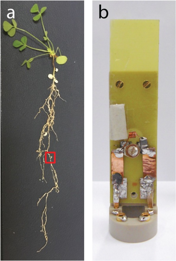
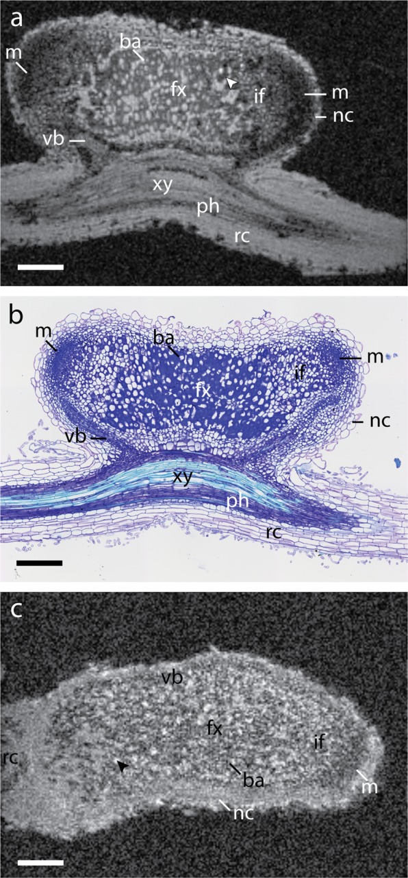
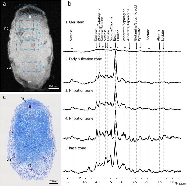
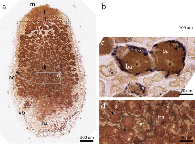
Interactions between plants and the soil's microbial & fungal flora are crucial for the health of soil ecosystems and food production. Microbe-plant interactions are difficult to investigate in situ due to their intertwined relationship involving morphology and metabolism. Here, we describe an approach to overcome this challenge by elucidating morphology and the metabolic profile of Medicago truncatula root nodules using Magnetic Resonance (MR) Microscopy, at the highest magnetic field strength (22.3 T) currently available for imaging. A home-built solenoid RF coil with an inner diameter of 1.5 mm was used to study individual root nodules. A 3D imaging sequence with an isotropic resolution of (7 μm)3 was able to resolve individual cells, and distinguish between cells infected with rhizobia and uninfected cells. Furthermore, we studied the metabolic profile of cells in different sections of the root nodule using localised MR spectroscopy and showed that several metabolites, including betaine, asparagine/aspartate and choline, have different concentrations across nodule zones. The metabolite spatial distribution was visualised using chemical shift imaging. Finally, we describe the technical challenges and outlook towards future in vivo MR microscopy of nodules and the plant root system.


Conflict of interest statement

The authors declare no competing interests.

Figures

Figure 1
Figure 1
Photograph of M. truncatula root system and custom-designed home-built microcoil. (a) Photograph of a representative M. truncatula plant (five weeks old, three weeks post inoculation). Typical root nodule indicated with a red square. Some white perlite beads are still visible along the root system. (b) Photograph of a home-built solenoid coil insert mounted on a Bruker insert holder. The solenoid inner diameter is 1.5 mm; further details are described in the materials and methods.
Figure 2
Figure 2
High-resolution MR imaging and optical microscopy of root nodules. (a) FLASH image of fixed root nodule at 7 × 7 × 7 µm3 resolution with an acquisition time of 34 h24 m. Individual cells containing bacteroids (ba) can be discerned by the dark rings inside the cells, where S. meliloti cells accumulate. The nodule exhibits a double meristem (m) on opposed directions with cells becoming smaller towards the meristem until individual cells can no longer be resolved. Air pockets appear as hypo-intense regions marked with an arrowhead. (b) Optical transmission microscopy of nodule section (thickness 5 µm) stained with Toluidine blue. (c) FLASH image of in situ root nodule at 7 × 7 × 7 µm3 resolution with an acquisition time of 34 h24 m. Though lower in signal-to-noise, both uninfected cells (black arrow) and infected cells (ba) can be discerned. Abbreviations: rc, root cortex; xy, xylem; ph, phloem; nc, nodule cortex; vb, vascular bundle; fx, fixation zone; if, infection zone; ba, bacteroid containing cells; m, meristem. Scale bars 200 µm.
Figure 3
Figure 3
Localised spectroscopy in in-situ root nodule reveals sugar differences in pre- and post-granule onset regions. (a) FLASH reference with PRESS Region of Interest (ROI), numbered one through five. Roman numerals indicate nodule zones: Meristem (I), Infection zone (II), Nitrogen fixation zone (III). Grey dotted boxes show ROI shift of Betaine due to Chemical Shift Displacement Error (CDSE). Pulse excitation was centred around 4.0 ppm. (b) PRESS spectra captured from Regions of Interest (ROI) shown in a, for Nodule Meristem (1), (early) Nitrogen fixation zone (2), Nitrogen fixation zone (3 & 4), Basal region (5). PRESS voxel sizes were 200 × 350 × 350 µm3. Spectra were aligned to the betaine peak at 3.3 ppm. Water peak region at 4.7 ppm has been omitted for ease of viewing. Intensities of the spectra have not been normalised, reflecting the strength of the signal recorded. This means that for the meristem little information can be discerned. Line broadening 10 Hz. (c) Co-registration of Optical Microscopy confirms the presence of bacteroids in the active nitrogen fixation region. Toluidine blue staining 10x magnification. Abbreviations: nc, nodule cortex; vb, vascular bundle; m, meristem; ra, root attachment area. Scale bars 200 µm.
Figure 4
Figure 4
Optical microscopy of root nodules reveals multiple starch distribution patterns. Coupes were stained with both Lugol’s solution and Toluidine blue. (a) A section immediately adjacent to that of Fig. 3c reveals starch distribution throughout the nodule. 10x magnification, scale bar 200 µm. A black dotted rectangle indicates the zone shown in (b) on an alternate section. A white rectangle indicates an enlargement seen in (d). 10x magnification, zoomed; scale bar 50 µm. (b) At the first layer of cells in zone III, starch is present as a band (s). A white rectangle indicates the area shown in (c). Alternate section, 20x Magnification, scale bar 100 µm. (c) Starch granules (s) are located near the periphery of infected cells. The vacuole (v) reappears in the fixation zone after disorganisation in the infection zone. (b). 40x Magnification, scale bar 20 µm. (d) A fine distribution of starch granules can be seen in Zone III contained in nitrogen fixating cells (black arrowhead) and uninfected cells (white arrowhead). Abbreviations: ra, root attachment site; nc, nodule cortex; vb, vascular bundles; m, meristem; s, starch granules; v, vacuole; ba, bacteroids.

References

    1. Doran JW, Zeiss MR. Soil health and sustainability: managing the biotic component of soil quality. Appl. Soil Ecol. 2000;15:3–11. doi: 10.1016/S0929-1393(00)00067-6. - DOI
    1. Smil, V. Nitrogen cycle and world food production. World Agric. 9–13 (2001).
    1. Smith BE. Nitrogenase reveals its inner secrets. Science. 2002;297:1654–1655. doi: 10.1126/science.1076659. - DOI - PubMed
    1. Suzaki, T., Yoro, E. & Kawaguchi, M. Leguminous Plants: Inventors of Root Nodules to Accommodate Symbiotic Bacteria. International Review of Cell and Molecular Biology316, (Elsevier Ltd, 2015). - PubMed
    1. Kim BH, Ramanan R, Cho DH, Oh HM, Kim HS. Role of Rhizobium, a plant growth promoting bacterium, in enhancing algal biomass through mutualistic interaction. Biomass and Bioenergy. 2014;69:95–105. doi: 10.1016/j.biombioe.2014.07.015. - DOI

Publication types