

Withanolide D Enhances Radiosensitivity of Human Cancer Cells by Inhibiting DNA Damage Non-homologous End Joining Repair Pathway
- PMID: 31970089
- PMCID: PMC6960174
- DOI: 10.3389/fonc.2019.01468
Withanolide D Enhances Radiosensitivity of Human Cancer Cells by Inhibiting DNA Damage Non-homologous End Joining Repair Pathway
Abstract
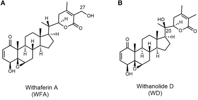
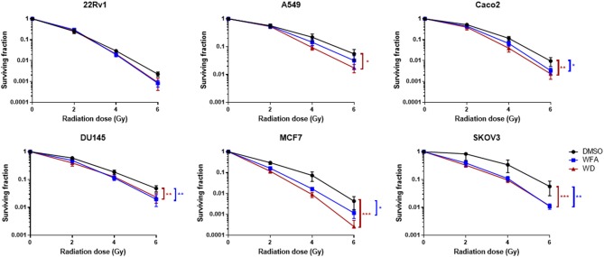
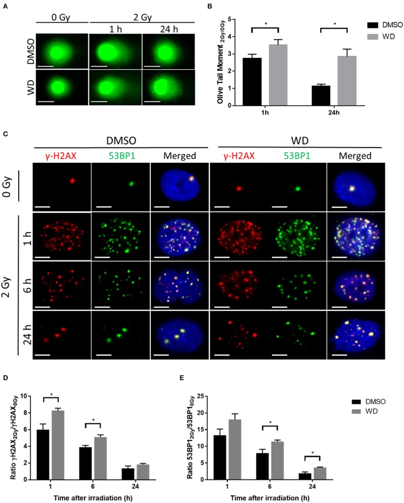
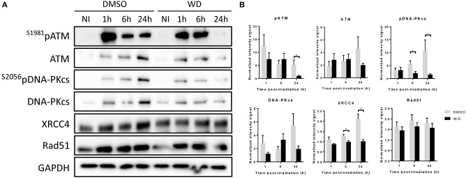
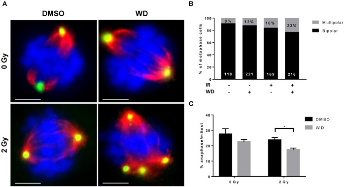
Along with surgery and chemotherapy, radiation therapy (RT) is an important modality in cancer treatment, and the development of radiosensitizers is a current key challenge in radiobiology to maximize RT efficiency. In this study, the radiosensitizing effect of a natural compound from the withanolide family, withanolide D (WD), was assessed. Clonogenic assays showed that a 1 h WD pretreatment (0.7 μM) before irradiation decreased the surviving fraction of several cancer cell lines. To determine the mechanisms by which WD achieved its radiosensitizing effect, we then assessed whether WD could promote radiation-induced DNA damages and inhibit double-strand breaks (DSBs) repair in SKOV3 cells. Comet and γH2AX/53BP1 foci formation assays confirmed that DSBs were higher between 1 and 24 h after 2 Gy-irradiation in WD-treated cells compared to vehicle-treated cells, suggesting that WD induced the persistence of radiation-induced DNA damages. Immunoblotting was then performed to investigate protein expression involved in DNA repair pathways. Interestingly, DNA-PKc, ATM, and their phosphorylated forms appeared to be inhibited 24 h post-irradiation in WD-treated samples. XRCC4 expression was also down-regulated while RAD51 expression did not change compared to vehicle-treated cells suggesting that only non-homologous end joining (NHEJ) pathways was inhibited by WD. Mitotic catastrophe (MC) was then investigated in SKOV3, a p53-deficient cell line, to assess the consequence of such inhibition. MC was induced after irradiation and was predominant in WD-treated samples as shown by the few numbers of cells pursuing into anaphase and the increased amount of bipolar metaphasic cells. Together, these data demonstrated that WD could be a promising radiosensitizer candidate for RT by inhibiting NHEJ pathway and promoting MC. Additional studies are required to better understand its efficiency and mechanism of action in more relevant clinical models.
Keywords: DNA damage repair; cancer; mitotic catastrophe; radiation; radiosensitizer; withanolide D.
Copyright © 2020 Lacombe, Cretignier, Meli, Wijeratne, Veuthey, Cuendet, Gunatilaka and Zenhausern.
Figures
Similar articles
-
Regulation of DNA repair in the absence of classical non-homologous end joining.DNA Repair (Amst). 2018 Aug;68:34-40. doi: 10.1016/j.dnarep.2018.06.001. Epub 2018 Jun 12. DNA Repair (Amst). 2018. PMID: 29929045
-
Regulation of ATM in DNA double strand break repair accounts for the radiosensitivity in human cells exposed to high linear energy transfer ionizing radiation.Mutat Res. 2009 Nov 2;670(1-2):15-23. doi: 10.1016/j.mrfmmm.2009.06.016. Epub 2009 Jul 5. Mutat Res. 2009. PMID: 19583974
-
The purine scaffold Hsp90 inhibitor PU-H71 sensitizes cancer cells to heavy ion radiation by inhibiting DNA repair by homologous recombination and non-homologous end joining.Radiother Oncol. 2016 Oct;121(1):162-168. doi: 10.1016/j.radonc.2016.08.029. Epub 2016 Sep 22. Radiother Oncol. 2016. PMID: 27666928 Free PMC article.
-
Mechanisms of DNA double strand break repair and chromosome aberration formation.Cytogenet Genome Res. 2004;104(1-4):14-20. doi: 10.1159/000077461. Cytogenet Genome Res. 2004. PMID: 15162010 Review.
-
DNA double strand break repair inhibition as a cause of heat radiosensitization: re-evaluation considering backup pathways of NHEJ.Int J Hyperthermia. 2008 Feb;24(1):17-29. doi: 10.1080/02656730701784782. Int J Hyperthermia. 2008. PMID: 18214766 Review.
Cited by
-
Photodynamic Therapy and Dietary Antioxidants: A Dual Strategy for Genome Stability and DNA Damage Repair.Cancer Med. 2025 Aug;14(15):e71032. doi: 10.1002/cam4.71032. Cancer Med. 2025. PMID: 40709614 Free PMC article. Review.
-
Preclinical Evidence of Withania somnifera and Cordyceps spp.: Neuroprotective Properties for the Management of Alzheimer's Disease.Int J Mol Sci. 2025 Jun 4;26(11):5403. doi: 10.3390/ijms26115403. Int J Mol Sci. 2025. PMID: 40508211 Free PMC article. Review.
-
Plant-Based Scaffolds Modify Cellular Response to Drug and Radiation Exposure Compared to Standard Cell Culture Models.Front Bioeng Biotechnol. 2020 Aug 7;8:932. doi: 10.3389/fbioe.2020.00932. eCollection 2020. Front Bioeng Biotechnol. 2020. PMID: 32850759 Free PMC article.
-
Natural Compounds That Target DNA Repair Pathways and Their Therapeutic Potential to Counteract Cancer Cells.Front Oncol. 2020 Nov 19;10:598174. doi: 10.3389/fonc.2020.598174. eCollection 2020. Front Oncol. 2020. PMID: 33330091 Free PMC article. Review.
-
LncRNA TMPO-AS1 Promotes Triple-Negative Breast Cancer by Sponging miR-383-5p to Trigger the LDHA Axis.Asian Pac J Cancer Prev. 2024 Aug 1;25(8):2929-2944. doi: 10.31557/APJCP.2024.25.8.2929. Asian Pac J Cancer Prev. 2024. PMID: 39205592 Free PMC article.
References
-
- Bharti VK, Malik JK, Gupta RC. Chapter 52 - Ashwagandha: multiple health benefits. In: Gupta RC, editor. Nutraceuticals. Boston: Academic Press; (2016). p. 717–33. 10.1016/B978-0-12-802147-7.00052-8 - DOI
LinkOut - more resources
Full Text Sources
Research Materials
Miscellaneous