

Can the wet suction technique change the efficacy of endoscopic ultrasound-guided fine-needle aspiration for diagnosing autoimmune pancreatitis type 1? A prospective single-arm study
- PMID: 31970173
- PMCID: PMC6962058
- DOI: 10.12998/wjcc.v8.i1.88
Can the wet suction technique change the efficacy of endoscopic ultrasound-guided fine-needle aspiration for diagnosing autoimmune pancreatitis type 1? A prospective single-arm study
Abstract
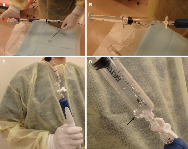
Background: Other than surgery, endoscopic ultrasound-guided fine-needle aspiration (EUS-FNA) is the only procedure for histologically diagnosing autoimmune pancreatitis (AIP). However, adequate specimens are difficult to obtain. Recently, more adequate specimens were reported to be obtained with EUS-FNA with a wet suction technique (WEST) than with conventional EUS-FNA.
Aim: To histologically diagnose AIP by EUS-FNA with a WEST.
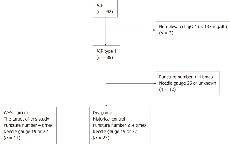
Methods: Eleven patients with possible type 1 AIP between February 2016 and August 2018 underwent EUS-FNA with a WEST (WEST group), with four punctures by 19 or 22 G needles. As a historical control, 23 type 1 AIP patients who underwent no fewer than four punctures with 19 or 22 G needles were enrolled (DRY group). Patient characteristics and histological findings were compared between the two groups.
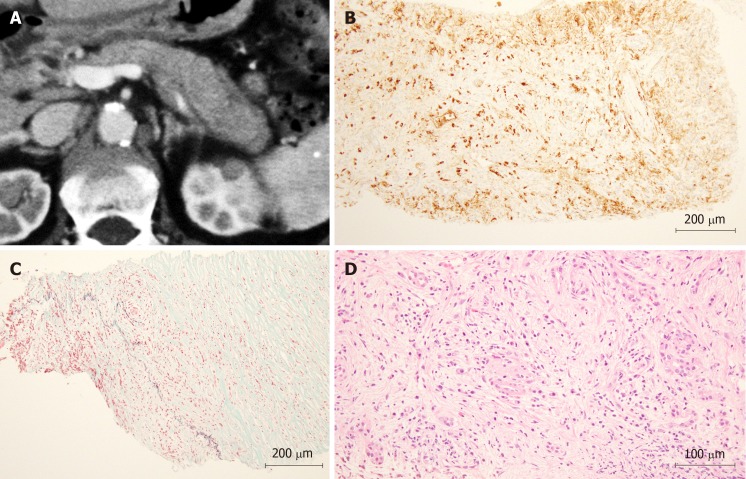
Results: Three histopathological factors according to the International Consensus Diagnostic Criteria were significantly greater in the WEST group than the DRY group [lymphoplasmacytic infiltrate without granulocytic infiltration: 9 (81.8%) vs 6 (26.1%), P = 0.003, storiform fibrosis: 5 (45.5%) vs 1 (4.3%), P = 0.008, abundant (> 10 cells/HPF) IgG4-positive cells: 7 (63.6%) vs 5 (21.7%), P = 0.026]. Level 1 or level 2 histopathological findings were observed more often in the WEST group than in the DRY group [8 (72.7%) vs 3 (13.0%), P = 0.001].
Conclusion: EUS-FNA with a WEST was more successful than standard EUS-FNA in histologically diagnosing AIP.
Keywords: Autoimmune pancreatitis; Endoscopic ultrasound-guided fine needle aspiration; Wet suction technique.
©The Author(s) 2020. Published by Baishideng Publishing Group Inc. All rights reserved.
Conflict of interest statement
Conflict-of-interest statement: The authors of this manuscript having no conflicts of interest to disclose.
Figures
Similar articles
-
The Role of EUS-Guided FNA and FNB in Autoimmune Pancreatitis.Diagnostics (Basel). 2021 Sep 9;11(9):1653. doi: 10.3390/diagnostics11091653. Diagnostics (Basel). 2021. PMID: 34573995 Free PMC article. Review.
-
The role of EUS-guided fine needle aspiration in autoimmune pancreatitis: a single center prospective study.Scand J Gastroenterol. 2018 Dec;53(12):1604-1610. doi: 10.1080/00365521.2018.1534137. Epub 2018 Nov 13. Scand J Gastroenterol. 2018. PMID: 30422724
-
Endoscopic ultrasound-guided fine-needle biopsy needle can facilitate histological diagnosis of type 1 autoimmune pancreatitis.J Hepatobiliary Pancreat Sci. 2025 Mar;32(3):238-245. doi: 10.1002/jhbp.12095. Epub 2024 Dec 6. J Hepatobiliary Pancreat Sci. 2025. PMID: 39639754
-
Diagnosis of autoimmune pancreatitis by EUS-guided FNA using a 22-gauge needle: a prospective multicenter study.Gastrointest Endosc. 2016 Nov;84(5):797-804.e1. doi: 10.1016/j.gie.2016.03.1511. Epub 2016 Apr 9. Gastrointest Endosc. 2016. PMID: 27068878
-
Role of endoscopy in the diagnosis of autoimmune pancreatitis and immunoglobulin G4-related sclerosing cholangitis.Dig Endosc. 2014 Sep;26(5):627-35. doi: 10.1111/den.12289. Epub 2014 Apr 8. Dig Endosc. 2014. PMID: 24712522 Review.
Cited by
-
Wet- versus dry-suction techniques for EUS-FNA of solid lesions: A systematic review and meta-analysis.Endosc Ultrasound. 2021 Sep-Oct;10(5):319-324. doi: 10.4103/EUS-D-20-00198. Endosc Ultrasound. 2021. PMID: 34259217 Free PMC article. Review.
-
The Role of EUS-Guided FNA and FNB in Autoimmune Pancreatitis.Diagnostics (Basel). 2021 Sep 9;11(9):1653. doi: 10.3390/diagnostics11091653. Diagnostics (Basel). 2021. PMID: 34573995 Free PMC article. Review.
-
A Pilot Randomized Crossover Trial of Wet Suction and Conventional Techniques of Endoscopic Ultrasound-Guided Fine-Needle Aspiration for Upper Gastrointestinal Subepithelial Lesions.Gastroenterol Res Pract. 2021 Mar 22;2021:4913107. doi: 10.1155/2021/4913107. eCollection 2021. Gastroenterol Res Pract. 2021. PMID: 33824658 Free PMC article.
-
Comparison of specimen quality among the standard suction, slow-pull, and wet suction techniques for EUS-FNA: A multicenter, prospective, randomized controlled trial.Endosc Ultrasound. 2022 Sep-Oct;11(5):393-400. doi: 10.4103/EUS-D-21-00163. Endosc Ultrasound. 2022. PMID: 36255027 Free PMC article.
-
Diagnostic yield of endoscopic ultrasound-guided tissue acquisition in autoimmune pancreatitis: a systematic review and meta-analysis.Endosc Int Open. 2021 Jan;9(1):E66-E75. doi: 10.1055/a-1293-7279. Epub 2021 Jan 1. Endosc Int Open. 2021. PMID: 33403238 Free PMC article.
References
-
- Yoshida K, Toki F, Takeuchi T, Watanabe S, Shiratori K, Hayashi N. Chronic pancreatitis caused by an autoimmune abnormality. Proposal of the concept of autoimmune pancreatitis. Dig Dis Sci. 1995;40:1561–1568. - PubMed
-
- Hamano H, Kawa S, Horiuchi A, Unno H, Furuya N, Akamatsu T, Fukushima M, Nikaido T, Nakayama K, Usuda N, Kiyosawa K. High serum IgG4 concentrations in patients with sclerosing pancreatitis. N Engl J Med. 2001;344:732–738. - PubMed
-
- Shimosegawa T, Chari ST, Frulloni L, Kamisawa T, Kawa S, Mino-Kenudson M, Kim MH, Klöppel G, Lerch MM, Löhr M, Notohara K, Okazaki K, Schneider A, Zhang L International Association of Pancreatology. International consensus diagnostic criteria for autoimmune pancreatitis: guidelines of the International Association of Pancreatology. Pancreas. 2011;40:352–358. - PubMed
-
- Ishikawa T, Itoh A, Kawashima H, Ohno E, Matsubara H, Itoh Y, Nakamura Y, Hiramatsu T, Nakamura M, Miyahara R, Ohmiya N, Goto H, Hirooka Y. Endoscopic ultrasound-guided fine needle aspiration in the differentiation of type 1 and type 2 autoimmune pancreatitis. World J Gastroenterol. 2012;18:3883–3888. - PMC - PubMed
-
- Imai K, Matsubayashi H, Fukutomi A, Uesaka K, Sasaki K, Ono H. Endoscopic ultrasonography-guided fine needle aspiration biopsy using 22-gauge needle in diagnosis of autoimmune pancreatitis. Dig Liver Dis. 2011;43:869–874. - PubMed
LinkOut - more resources
Full Text Sources
Research Materials
Miscellaneous
