. 2019 Dec 14;7(1):70-77.
doi: 10.1002/mdc3.12873. eCollection 2020 Jan.

Hyperphosphorylated Tau, Increased Adenylate Cyclase 5 (ADCY5) Immunoreactivity, but No Neuronal Loss in ADCY5-Dyskinesia

Affiliations

Hyperphosphorylated Tau, Increased Adenylate Cyclase 5 (ADCY5) Immunoreactivity, but No Neuronal Loss in ADCY5-Dyskinesia

Dong-Hui Chen et al. Mov Disord Clin Pract. .

Abstract

Background: Adenylate cyclase 5 (ADCY5)-related dyskinesia is a childhood-onset movement disorder. Manifestations vary in frequency and severity and may include chorea, tremor, dystonia, facial twitches, myoclonus, axial hypotonia, and limb hypertonia. Psychosis is likely part of the broader spectrum. ADCY5 is widely expressed in the brain, especially in the striatum. Previous reports of brain autopsies of 2 subjects with likely ADCY5-dyskinesia were limited by the absence of a molecular diagnosis. In 1 case, normal gross pathology was reported. In the other case, ADCY5 expression was not examined and neuropathological findings were confounded by age and comorbidities.

Objectives: To examine ADCY5 expression and neuropathological changes in ADCY5-dyskinesia.

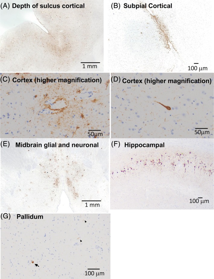
Methods: An extensive brain autopsy, including immunohistochemical analyses with antibodies to paired helical filament tau, α-synuclein, amyloid-β, microtubule-associated protein 2, and ADCY5, was performed.

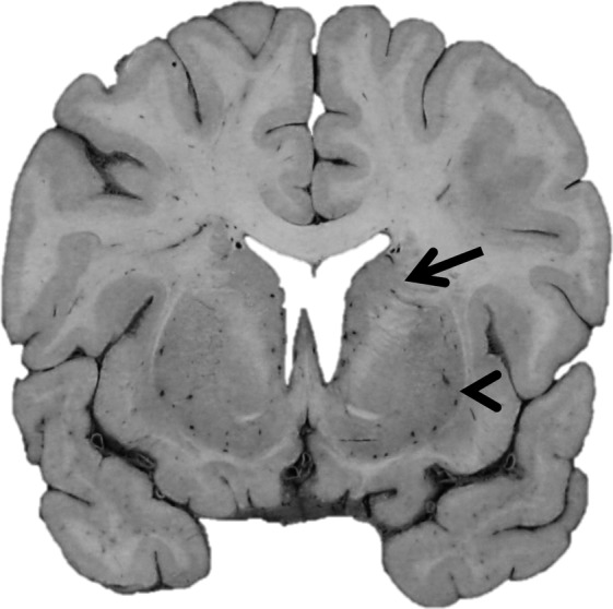
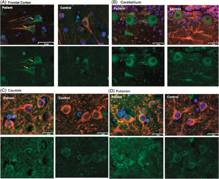
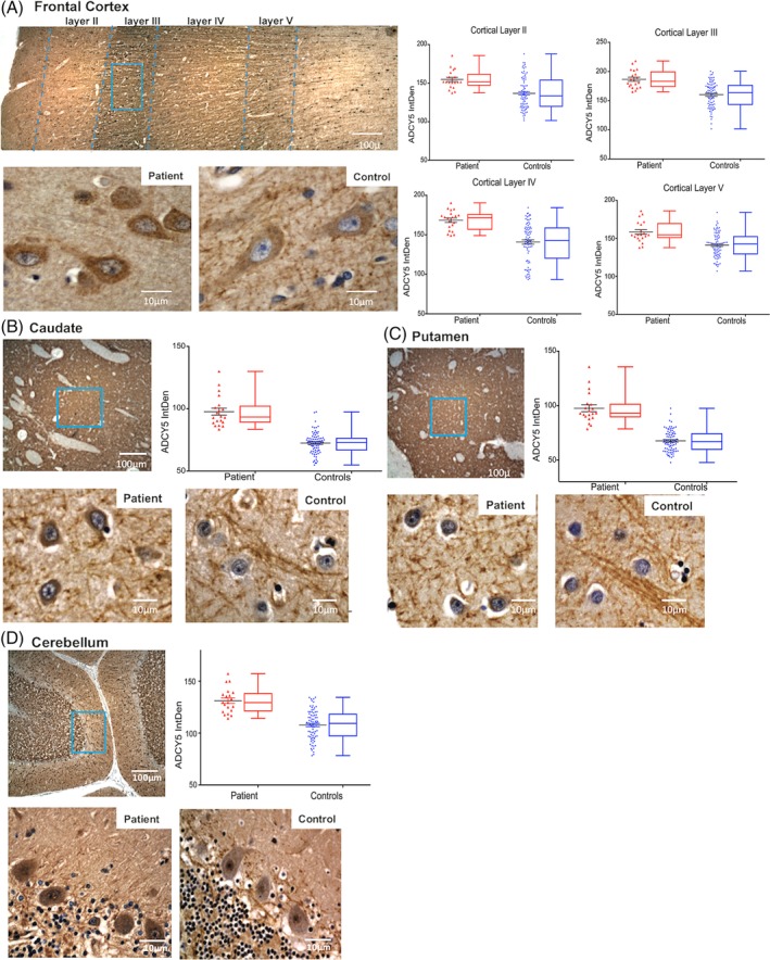
Results: The patient, with a p.M1029K ADCY5 variant, had severe dyskinesias from early childhood, later recurrent episodes of psychosis, and died at age 46. Gross pathology was unremarkable, but we detected increased immunoreactivity for ADCY5 in neurons in multiple brain regions. Despite no history of brain trauma to suggest chronic traumatic encephalopathy, we found tau deposits in the deep cortical sulci, midbrain, and hippocampus with minimal amyloid pathology and no Lewy bodies.

Conclusions: We present the first brain autopsy findings in a molecularly proven case of ADCY5-dyskinesia, showing increased ADCY5 immunoreactivity in neurons and evidence of tau deposition. Additional patients will need to be studied to determine whether increased immunoreactivity for ADCY5 is a signature for ADCY5-dyskinesia and whether this disease has a tauopathy component.

Keywords: ADCY5‐dyskinesia; neurogenetics; neuropathology; tau.


Conflict of interest statement

This work was supported by grants R01NS069719 and P50AG005136 from the National Institutes of Health, Merit Review Award Number 101 CX001702 from the United States Department of Veterans Affairs, and the Nancy and Buster Alvord Endowment. The authors report no conflicts of interest.

Figures

Figure 1
Figure 1
Gross pathology in a patient with ADCY5‐dyskinesia. Coronal section of the brain at the level of the anterior commissure demonstrating well‐formed caudate nucleus (arrow) and putamen (arrowhead) as well as no significant cortical atrophy or ventriculomegaly.
Figure 2
Figure 2
Microscopic neuropathology in a patient with ADCY5‐dyskinesia. PHF‐tau immunostained slides demonstrate patchy, but widespread, pTau pathology. (A) Sections of neocortex demonstrate neurofibrillary tangles along gyri as well as at the depths of sulci. (B) Cortical tau also consisted of subpial, astroglial tau inclusions. (C) Perivascular glial and neuritic tau pathology in the cortex. (D) High magnification of a neurofibrillary tangle. (E) Section of midbrain shows glial and neuronal tau in periaqueductal gray matter. (F) Section of hippocampus demonstrates neurofibrillary tangles in the CA1 subfield and (G) scattered tau neurites (arrowheads) and pretangles (arrow) of uncertain significance in the pallidum.
Figure 3
Figure 3
Expression and localization of ADCY5 in multiple brain areas in a patient with ADCY5‐dyskinesia. Double immunofluorescence staining of ADCY5 (in green) and the mature‐neuron marker, MAP2 (in red), in a patient with an ADCY5 p.M1029K missense variant and four age‐ and sex‐matched controls (1 representative control is shown). Nuclei are stained blue by DAPI. Multiple regions were evaluated: frontal cortex (A), cerebellum (B), caudate (C), and putamen (D). In each panel, top images show merged ADCY5 and MAP2, and the lower ones show ADCY5 only. Merged ADCY5 and MAP2 images show presence of ADCY5 in MAP2‐positive neurons, and not in MAP2‐negative cells, in all tested brain regions in the patient. Increased cytoplasmic granularity of ADCY5 is visible in the patient's cells (arrows in yellow), not seen with MAP2 staining only. Size bar: (A) and (B), 32 μm; (C) and (D), 16 μm.
Figure 4
Figure 4
Quantitative assessment of ADCY5 staining intensity in multiple brain regions in the patient with ADCY5‐dyskinesia. DAB chromogen IHC staining for ADCY5 in the patient and 4 age‐ and sex‐matched controls. In panel (A), the top image is full thickness of gray matter in the frontal cortex from the patient showing layers I through VI. Below, the higher‐magnification images show representative sections from that area framed in blue from the patient (left) and a control (right) stained for ADCY5. Similarly, panels (B), (C), and (D) are caudate, putamen, and cerebellum, respectively. For each area, the scatter and box‐whiskers plots display quantitative measurements of the DAB chromogen intensity of ADCY5 in 20 randomly selected cells in each subject. Results from the patient (data from 20 cells) are labeled in red, and results from 4 controls (combined data from a total of 80 cells) are grouped and labeled in blue. The results demonstrate increased ADCY5 staining intensity in the patient in all brain regions studied.

References

    1. Chen DH, Meneret A, Friedman JR, et al. ADCY5‐related dyskinesia: broader spectrum and genotype‐phenotype correlations. Neurology 2015;85:2026–2035. - PMC - PubMed
    1. Chang FC, Westenberger A, Dale RC, et al. Phenotypic insights into ADCY5‐associated disease. Mov Disord 2016;31:1033–1040. - PMC - PubMed
    1. Vijiaratnam N, Newby R, Kempster PA. Depression and psychosis in ADCY5‐related dyskinesia‐part of the phenotypic spectrum? J Clin Neurosci 2018;57:167–168. - PubMed
    1. Barrett MJ, Williams ES, Chambers C, Dhamija R. Autosomal recessive inheritance of ADCY5‐related generalized dystonia and myoclonus. Neurol Genet 2017;3:193. - PMC - PubMed
    1. Bohlega SA, Abou‐Al‐Shaar H, AlDakheel A, et al. Autosomal recessive ADCY5‐related dystonia and myoclonus: expanding the genetic spectrum of ADCY5‐related movement disorders. Parkinsonism Relat Disord 2019;64:145–149. - PubMed

LinkOut - more resources