

doi: 10.1002/mdc3.12846.
eCollection 2020 Jan.
Parkinsonism with Normal Dopaminergic Presynaptic Terminals in Cerebrotendinous Xanthomatosis
Affiliations
- PMID: 31970228
- PMCID: PMC6962677
- DOI: 10.1002/mdc3.12846
Item in Clipboard
Parkinsonism with Normal Dopaminergic Presynaptic Terminals in Cerebrotendinous Xanthomatosis
Mov Disord Clin Pract.
.
No abstract available
Keywords: cerebrotendinous xanthomatosis; dopaminergic presynaptic terminals; iron deposition; parkinsonian syndrome.
Conflict of interest statement
This study was funded by the Beijing Municipal Administration of Hospitals’ Mission Plan (Grant SML20150803), the Beijing Municipal Science & Technology Commission (Grant Z171100000117013), and the National Key R&D Program of China (Grants 2017YFC0840105 and 2018YFC1312000). The authors declare that there are no conflicts of interest relevant to this work.
Figures
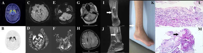
Imaging results. (A,B) [18F]‐9‐fluoropropyl‐(+)‐dihydrotetrabenazine positron emission tomography revealed normal striatal uptake of [18F]‐9‐fluoropropyl‐(+)‐dihydrotetrabenazine (arrows). Susceptibility‐weighted imaging (C,E) and quantitative susceptibility mapping (D,F) revealed iron deposition within several brain regions (arrows). T1‐weighted brain magnetic resonance imaging revealed abnormal, symmetric signals in the cerebellar dentate nucleus (G) as well as white matter degeneration (H). T1‐weighted right‐ankle magnetic resonance imaging revealed fusiform thickening (I,J; arrows). (K) Photograph of the enlarged Achilles tendon. Hematoxylin‐and‐eosin stained biopsy specimen of the Achilles tendon shows multinucleated giant cells (L; arrow), xanthoma cells (M; thick arrow), and dispersed lipid crystal clefts (M; thin arrow). Magnifications: G, 200×; H, 100×.
References
-
- Stelten BML, Warrenburg BPCVD, Wevers RA, Verrips A. Movement disorders in cerebrotendinous xanthomatosis. Parkinsonism Relat Disord 2019;58:12–16. - PubMed
-
- Makary MS, Kisanuki YY, Amin NN, Slone HW. Teaching neuroimages: cerebrotendinous xanthomatosis: a rare treatable adult‐onset lipid storage disease. Neurology 2018;90(7):e637–e638. - PubMed
-
- Brar S, Henderson D, Schenck J, Zimmerman EA. Iron accumulation in the substantia nigra of patients with Alzheimer disease and parkinsonism. JAMA Neurology 2009;66(3):371–374. - PubMed
-
- Hider RC, Longo DL, Hoffbrand AV. The role of deferiprone in iron chelation. N Engl J Med 2018;379(22):2140–2150. - PubMed
LinkOut - more resources
Full Text Sources
