. 2020 Feb 12;142(6):2704-2708.
doi: 10.1021/jacs.9b10984. Epub 2020 Jan 30.

Imidazole-Imidazole Hydrogen Bonding in the pH-Sensing Histidine Side Chains of Influenza A M2

Affiliations

Imidazole-Imidazole Hydrogen Bonding in the pH-Sensing Histidine Side Chains of Influenza A M2

Kumar Tekwani Movellan et al. J Am Chem Soc. .

Abstract

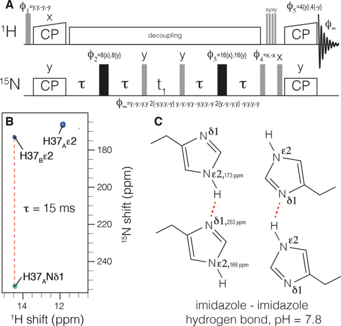
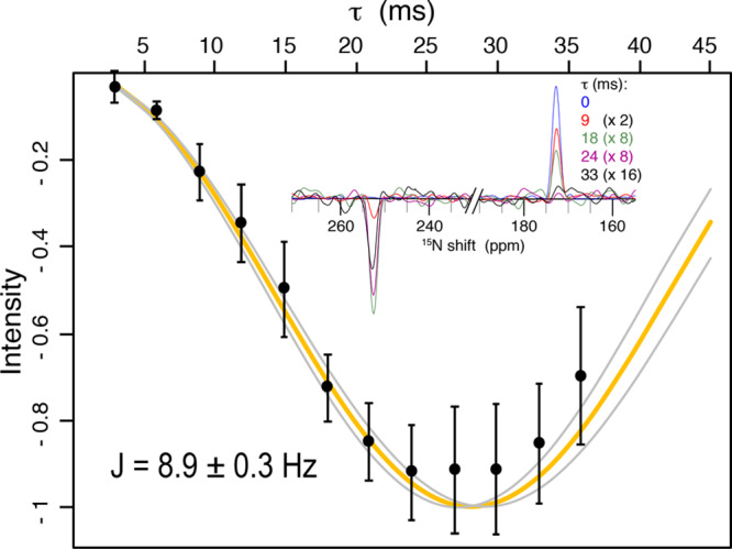
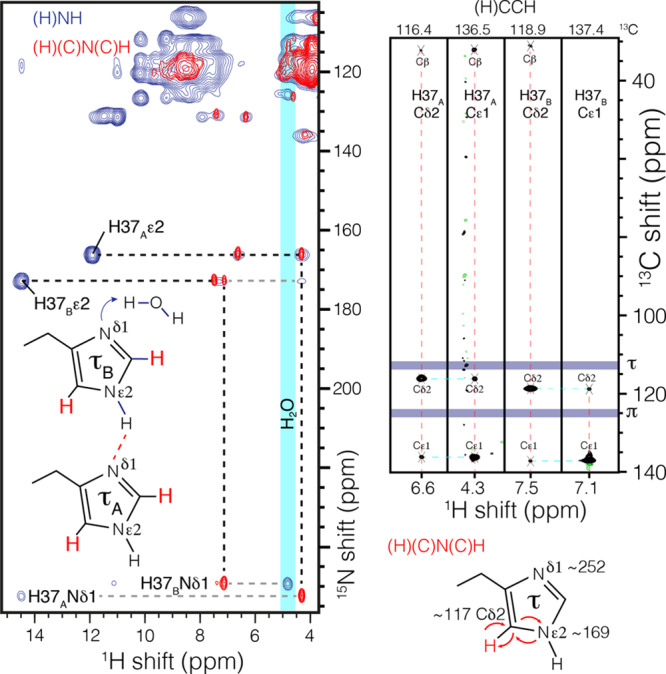
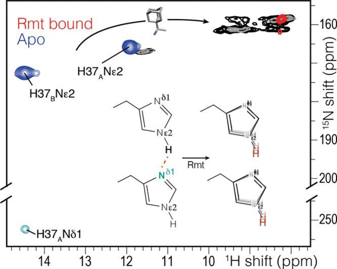
The arrangement of histidine side chains in influenza A M2 tetramer determines their pKa values, which define pH-controlled proton conduction critical to the virus lifecycle. Both water-associated and hydrogen-bonded imidazole-imidazolium histidine quaternary structures have been proposed, based on crystal structures and NMR chemical shifts, respectively. Here we show, using the conduction domain construct of M2 in lipid bilayers, that the imidazole rings are hydrogen bonded even at a pH of 7.8 in the neutral charge state. An intermolecular 8.9 ± 0.3 Hz 2hJNN hydrogen bond is observed between H37 Nε and Nδ recorded in a fully protonated sample with 100 kHz magic-angle spinning. This interaction could not be detected in the drug-bound sample.


Conflict of interest statement

The authors declare no competing financial interest.

Figures

Figure 1
Figure 1
Measurement of 2hJNHN hydrogen bonding in H37 imidazole dimers within influenza M2. The pulse sequence is shown in (a). Cross-polarization (CP) is used to establish 15N polarization. A homonuclear out-and-back INEPT period follows to record the chemical shift of the J-coupled nitrogen. Following water suppression, CP returns signal to protons for detection. The spectrum in (b) was recorded with a τ of 15 ms and clearly shows a negative peak indicative of an intermolecular J-coupling and a C2 symmetric tetramer at H37, as shown schematically in (c).
Figure 2
Figure 2
Quantification of the intermolecular 2hJNHNJ-coupling. In the inset, slices of the 2D spectrum at the proton frequency of 14.5 are shown for the indicated mixing times. The experimental data (points) are shown with 2σ error bars accounting for random spectral noise. Relaxation was accounted for by dividing each intensity at 254 ppm by the total signal magnitude of the slice. The best fit (orange) resulted in a coupling strength of 8.9 ± 0.3 Hz. The curves in gray indicate the error at twice the standard deviation, σ, as estimated with a Monte Carlo approach and considering random spectral noise. The first point was acquired with 8 scans (1.5 h), while the last point required 128 scans (26 h) due to transverse relaxation (see Figure S1).
Figure 3
Figure 3
Histidine–water contact and assignment of the H37 tautomer state. In blue, an NH correlation spectrum shows magnetization transfer from a nonprotonated imidazole nitrogen at ∼250 ppm to water (4.85 ppm) using 6 ms of CP. In red, the nitrogen and carbon resonances are assigned by out-and-back one-bond CP transfer (H)(C)N(C)H. The Hδ2 is correlated only with Nε2 in this magnetization transfer scheme, which resonates at ∼170 ppm and establishes that all histidine residues in the channel are in the τ tautomer. Magnetization transfers are indicated by curved arrows. The δ and ε carbon assignments were confirmed in an RFDR-based (H)CCH spectrum (black).
Figure 4
Figure 4
Chemical shift changes in the histidine side chain upon addition of the drug rimantadine (Rmt) using the pulse sequence of Figure 1 with a τ of 6 ms (blue, red) and (H)NH spectra with 25 ms (gray) or 200 ms (black) of 15N exchange during the water suppression. A 3–5 ppm change is observed in the drug-bound spectrum (red), and no 2hJNHNJ-coupling was observed. Instead, the imidazole NH peaks are broadened, and the peaks at 9 ppm are in exchange. The (H)NH spectra were acquired at 250 K and 80 kHz MAS to reduce the temperature by ∼10 °C to slow exchange.

References

    1. Kochendoerfer G. G.; Salom D.; Lear J. D.; Wilk-Orescan R.; Kent S. B.; DeGrado W. F. Total chemical synthesis of the integral membrane protein influenza A virus M2: role of its C-terminal domain in tetramer assembly. Biochemistry 1999, 38 (37), 11905–13. 10.1021/bi990720m. - DOI - PubMed
    1. Sakaguchi T.; Tu Q.; Pinto L. H.; Lamb R. A. The active oligomeric state of the minimalistic influenza virus M2 ion channel is a tetramer. Proc. Natl. Acad. Sci. U. S. A. 1997, 94 (10), 5000–5. 10.1073/pnas.94.10.5000. - DOI - PMC - PubMed
    1. Sugrue R. J.; Hay A. J. Structural characteristics of the M2 protein of influenza A viruses: evidence that it forms a tetrameric channel. Virology 1991, 180 (2), 617–24. 10.1016/0042-6822(91)90075-M. - DOI - PMC - PubMed
    1. Sugrue R. J.; Bahadur G.; Zambon M. C.; Hall-Smith M.; Douglas A. R.; Hay A. J. Specific structural alteration of the influenza haemagglutinin by amantadine. EMBO J. 1990, 9 (11), 3469–76. 10.1002/j.1460-2075.1990.tb07555.x. - DOI - PMC - PubMed
    1. Colvin M. T.; Andreas L. B.; Chou J. J.; Griffin R. G. Proton association constants of His 37 in the Influenza-A M218–60 dimer-of-dimers. Biochemistry 2014, 53 (38), 5987–94. 10.1021/bi5005393. - DOI - PMC - PubMed

Publication types

MeSH terms