Review
. 2020 Apr:63:57-67.
doi: 10.1016/j.ceb.2019.12.007. Epub 2020 Jan 20.

Novel roles of phosphoinositides in signaling, lipid transport, and disease

Affiliations
Review

Novel roles of phosphoinositides in signaling, lipid transport, and disease

Gerald R V Hammond et al. Curr Opin Cell Biol. 2020 Apr.

Abstract

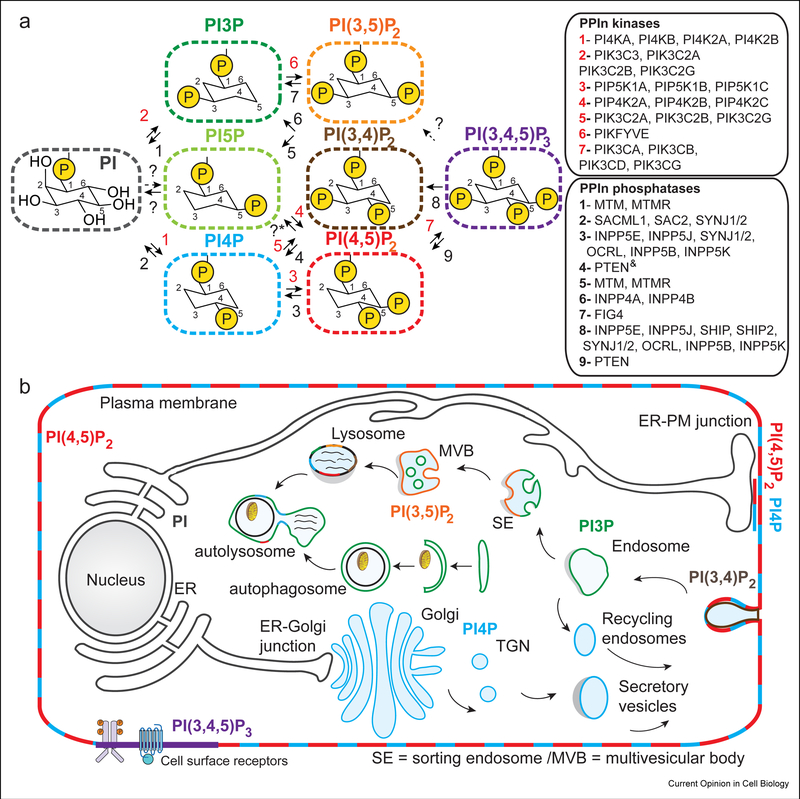
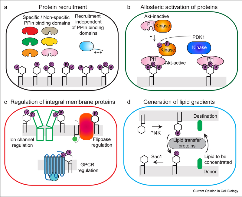
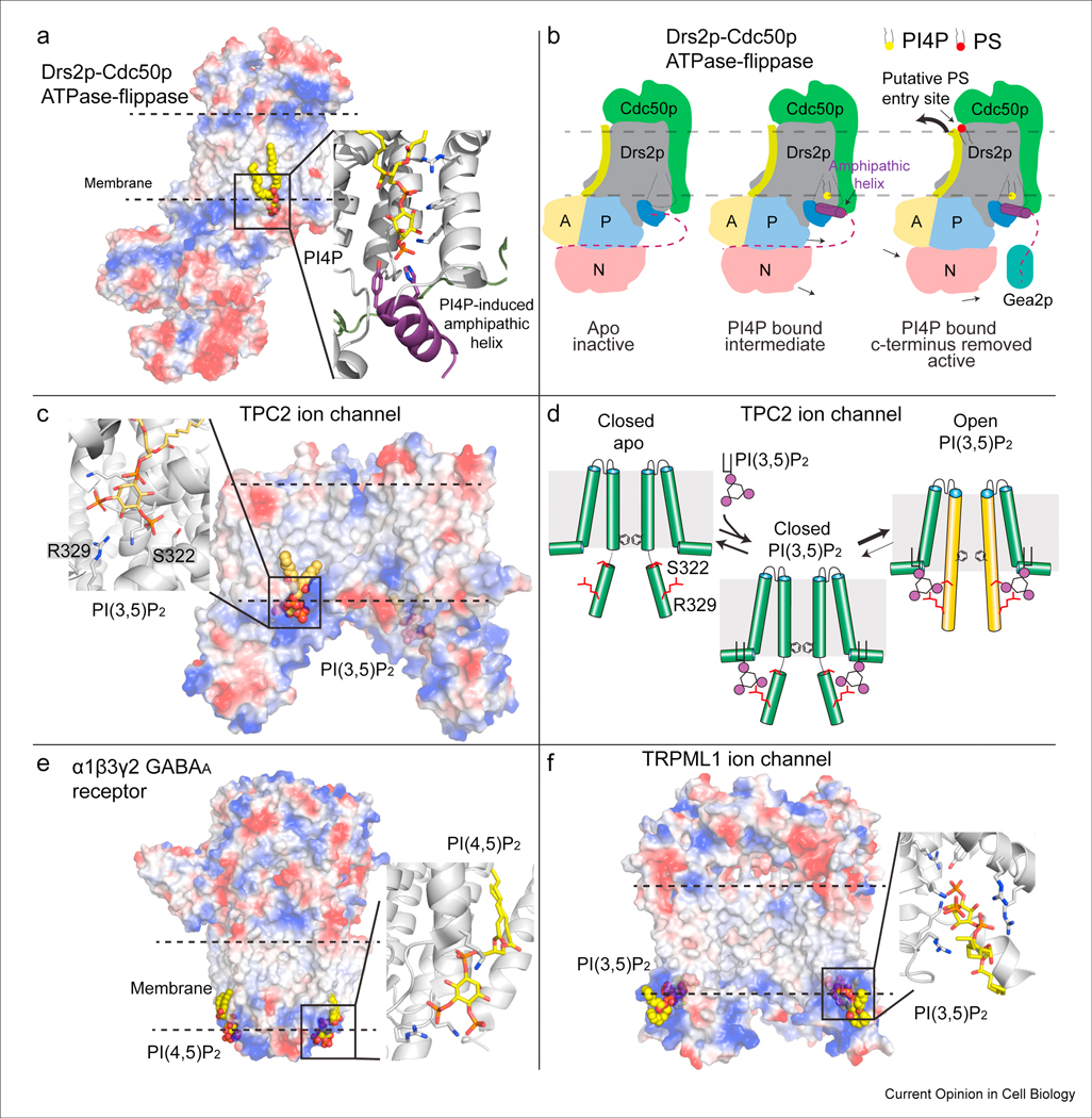
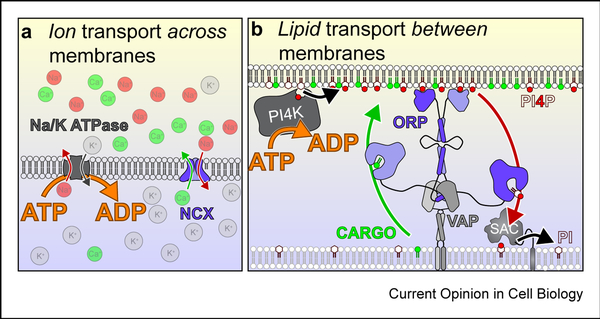
Phosphoinositides (PPIns) are lipid signaling molecules that act as master regulators of cellular signaling. Recent studies have revealed novel roles of PPIns in myriad cellular processes and multiple human diseases mediated by misregulation of PPIn signaling. This review will present a timely summary of recent discoveries in PPIn biology, specifically their role in regulating unexpected signaling pathways, modification of signaling outcomes downstream of integral membrane proteins, and novel roles in lipid transport. This has revealed new roles of PPIns in regulating membrane trafficking, immunity, cell polarity, and response to extracellular signals. A specific focus will be on novel opportunities to target PPIn metabolism for treatment of human diseases, including cancer, pathogen infection, developmental disorders, and immune disorders.

Keywords: Flippases; GPCR; Ion channels; Lipid kinases; Lipid signaling; Lipid transfer proteins; Membrane contact sites; Membrane trafficking; PI3P; PI4KB; PI4P; PIK3CA; PIP2; PIP3; Phosphatidylinositol; Phosphoinositide kinases; Phosphoinositides.


Conflict of interest statement

Conflict of interest statement Nothing declared.

Figures

Figure 1:
Figure 1:. Identity and cellular localization of phosphoinositides in cells
A. Phosphoinositides are composed of two acyl chains attached to a glycerol backbone, with a myo-inositol headgroup. There are a total of seven different phosphoinositide species that can be generated downstream of the precursor phosphatidylinositol through phosphorylation of the hydroxyls on the inositol headgroup. These include phosphatidylinositol 3-phosphate (PI3P), phosphatidylinositol 4-phosphate (PI4P), phosphatidylinositol 5-phosphate (PI5P), phosphatidylinositol 3,4 bis-phosphate (PI(3,4)P2, phosphatidylinositol 3,5 bis-phosphate (PI(3,4)P2, phosphatidylinositol 3,4 bis-phosphate (PI(4,5)P2, and phosphatidylinositol 3,4,5 tris-phosphate (PI(3,4,5)P2 (referred to as PIP3). The different human lipid kinases and phosphatases that generate them are indicated in the legend according to the numbers. Phosphoinositide conversion reactions that are not fully established are marked with a ? sign. *The conversion of PI4,5P2 to PI5P has been implicated to involve the PIP4P1/PIP4P2 genes (TMEM55A/TMTM55B proteins), however, recent work indicates the biological activity of these proteins is not driven through PI4,5P2 phosphatase activity [88]. The & sign indicates reactions that have only recently been identified [89]. B. The generation of phosphoinositides are master regulators of temporal and spatial localization of cellular signaling and membrane trafficking events, with their location tightly restricted through the action of the lipid kinases and phosphatases that generate them. They play key roles in secretion from the Golgi, endocytosis, and endo-lysosomal trafficking of membranes [90].
Figure 2:
Figure 2:. Phosphoinositides roles in protein recruitment/allosteric activation, modulation of integral membrane proteins, and lipid transport
A. Role of Phosphoinositides in the recruitment of proteins to specific intracellular locations. Many PPIn binding domains have been identified, although many of these domains have varying levels of specificity, and also frequently require coincidence detection of other signals (including both additional lipid and protein binding partners). Phosphoinositides can also regulate protein recruitment outside of lipid binding domains, including polybasic stretches, and non-canonical lipid binding sites. B. Roles of phosphoinositides in allosteric activation of signaling enzymes. Example of the allosteric activation of the pro-growth kinase Akt (PKB) downstream of PIP3, where PIP3 binding to the PH domain disrupts an inhibitory PH-kinase interface, followed by PIP3 activated phosphorylation of Akt by phosphoinositide dependent kinase 1 (PDK1). C. Phosphoinositides are key regulators of integral membrane proteins, including ion channels, G-protein coupled receptors, and lipid scramblases, flippases and floppases. Phosphoinositides can regulate integral membrane function through allosteric conformational changes and/or through modulating their coupling to protein binding partners. D. Phosphoinositides can mediate the transport of lipids against their concentration gradient through the coordinated action of lipid kinases, phosphatases, and lipid transport proteins (LTPs).
Figure 3:
Figure 3:. Structural basis for the regulation of integral membrane proteins by PPIns
A. Cryo-EM structure of the yeast dimeric complex of the ATPase flippase Drs2pCdc50p [29]. The protein complex is shown as a surface representation, highlighting charged pockets that mediate lipid binding. The amphipathic helix that binds PI4P is colored purple, with the residues that bind specifically to PI4P shown as sticks. B. A cartoon schematic of the conformational changes that occur during PI4P binding in the flippase catalytic cycle [29,91]. The Cdc50p protein is shown in green, with the Drs2p protein colored according to its domains, with the A, P, and N domains colored in yellow, blue and red respectively. The inhibitory c-terminus of Drs2p is shown as a dotted line, which is attached to the amphipathic helix that forms upon PI4P binding. The coordinated binding of PI4P and disruption of the c-terminal inhibitory interaction through binding to the Arf-GEF Gea2p leads to an allosteric conformational change in TM2 (colored lime) that opens a putative PS lipid binding pocket, allowing for lipid transfer. C. Cryo-EM structure of the open active form of the human Na2+ selective two pore channel (TPC2) bound to PI3,5P2 [35]. The protein complex is shown as a surface representation, highlighting charged pockets that mediate lipid binding. The specific residues that mediate phosphoinositide binding are shown as sticks. The Arg and Ser residues that interact with PI3,5P2 are labeled. D. Cartoon schematic of the molecular mechanism of how PI3,5P2 mediates ion channel opening (only the 6 TM-I domain is shown for simplicity). Binding of PI3,5P2 biases the equilibrium to the open conformation through allosteric conformational changes in the IS6 helix (colored in orange in the open conformation). This helix contains Ser-322 and Arg-329 which interact with PI3,5P2, and putatively bias the channel towards an open conformation. E. Cryo-EM structure of the type A γ-aminobutyric acid (GABAA) pentameric ligand gated ion channel bound to PI(4,5)P2 [30]. The protein complex is shown as a surface representation, highlighting charged pockets that mediate lipid binding. The specific residues that mediate phosphoinositide binding are shown as sticks. F. Cryo-EM structure of the Transient receptor potential mucolipin 1 (TRPML1) ion channel bound to PI(3,5)P2 [23]. The protein complex is shown as a surface representation, highlighting charged pockets that mediate lipid binding. The specific residues that mediate phosphoinositide binding are shown as sticks.
Figure 4:
Figure 4:. PPIn synthesis powers lipid transfer
Principles of counter ion (A) and counter-lipid (B) transport. In both cases, transport of a cargo (green) is powered by flow of the counter-molecule (red) down its concentration gradient. Ultimately, the chemical gradient of counter-molecule is established via ATP hydrolysis. This concept mirrors the textbook example of exchangers in ion homeostasis. For example, sodium and calcium ions are both maintained at low cytosolic concentrations relative to the extracellular milieu. The sodium calcium exchanger (NCX) helps maintain low cytosolic calcium concentrations in the absence of ATP hydrolysis, by exchanging cytosolic calcium ions for extracellular sodium ions. Thus calcium moves against its electrochemical gradient (out of cells), but powered by the flow of sodium down its own gradient (into cells). Ultimately, ATP powers this cycle through the sodium-potassium ATPases that actively pump sodium out of cells and maintains the gradient. The same has been proposed for lipid transfer proteins of the ORP family: These can exchange a sterol or phospholipid that is synthesized in the ER for PI4P in another membrane [10]. This membrane is anchored to the ER by the ORP protein itself, usually by PI4P through a PH domain at one end and through an ER anchor (or interaction with the ER receptor VAPa/b) at the other. The ORD domain then exchanges PI4P and cargo lipid. Hydrolysis of the PI4P in the ER by the SAC1 lipid phosphatase ensures vectoral transfer of PI4P, which is unavailable for the return step. Thus the cargo lipid is moved instead, against its own concentration gradient. Ultimately, ATP hydrolysis powers this cycle through the PI 4-kinases that maintains high PI4P levels in the non-ER membrane. Although this is an apparently novel concept relative to the protein recruitment and activation mechanisms traditionally associated with PPIn signaling, there is a unifying theme: In both cases, PPIn synthesis couples the energy of ATP hydrolysis to the nonequilibrium acquisition of lipid or protein molecules (or their activity) in restricted membrane compartments. This is an essential function for multi-organelle eukaryotic cells.

References

    1. Balla T: Phosphoinositides: tiny lipids with giant impact on cell regulation. Physiol. Rev. 2013, 93:1019–1137. - PMC - PubMed
    1. Goncalves MD, Cantley LC: Phosphatidylinositol 3-Kinase, Growth Disorders, and Cancer. N. Engl. J. Med. 2018, 379:2052–2062.

      * An excellent review on the involvement of PI3Ks in pathological conditions, and novel therapeutic strategies to target them in disease,

    1. Burke JE: Structural Basis for Regulation of Phosphoinositide Kinases and Their Involvement in Human Disease. Mol. Cell 2018, 71:653–673. - PubMed
    1. Behjati S, Tarpey PS, Sheldon H, Martincorena I, Van Loo P, Gundem G, Wedge DC, Ramakrishna M, Cooke SL, Pillay N, et al.: Recurrent PTPRB and PLCG1 mutations in angiosarcoma. Nat. Genet. 2014, 46:376–379. - PMC - PubMed
    1. Ketel K, Krauss M, Nicot A-S, Puchkov D, Wieffer M, Müller R, Subramanian D, Schultz C, Laporte J, Haucke V: A phosphoinositide conversion mechanism for exit from endosomes. Nature 2016, 529:408–412. - PubMed

Publication types

Substances