. 2020 Jan 21;9(2):265.
doi: 10.3390/cells9020265.

Meniscus Matrix Remodeling in Response to Compressive Forces in Dogs

Affiliations

Meniscus Matrix Remodeling in Response to Compressive Forces in Dogs

Umberto Polito et al. Cells. .

Abstract

Joint motion and postnatal stress of weight bearing are the principal factors that determine the phenotypical and architectural changes that characterize the maturation process of the meniscus. In this study, the effect of compressive forces on the meniscus will be evaluated in a litter of 12 Dobermann Pinschers, of approximately 2 months of age, euthanized as affected by the quadriceps contracture muscle syndrome of a single limb focusing on extracellular matrix remodeling and cell-extracellular matrix interaction (i.e., meniscal cells maturation, collagen fibers typology and arrangement). The affected limbs were considered as models of continuous compression while the physiologic loaded limbs were considered as controls. The results of this study suggest that a compressive continuous force, applied to the native meniscal cells, triggers an early maturation of the cellular phenotype, at the expense of the proper organization of collagen fibers. Nevertheless, an application of a compressive force could be useful in the engineering process of meniscal tissue in order to induce a faster achievement of the mature cellular phenotype and, consequently, the earlier production of the fundamental extracellular matrix (ECM), in order to improve cellular viability and adhesion of the cells within a hypothetical synthetic scaffold.

Keywords: GAGs; Young’s compressive elastic modulus; cell–extracellular matrix interaction; compression; dog.; extracellular matrix remodeling; meniscus; proteoglycans and glycosaminoglycans.


Conflict of interest statement

The authors declare no conflict of interest.

Figures
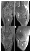
Figure 1
Figure 1
MR acquisition of a quadriceps-contracture-syndrome-affected knee and a healthy one. Coronal (A,C) and sagittal (B,D) reformat of 3D T1-weighted MR acquisition. A,B: right contracted limb; C,D: left noncontracted limb. The different morphology of menisci is clearly seen (white arrow, lateral meniscus body; black arrow, medial meniscus body; F, femur; T, tibia).
Figure 2
Figure 2
On the left, study design. The preparation of the meniscal samples in base of the type of technique performed is shown on the top. AH: anterior horn; BO: body; PH; posterior horn. Original picture by U.P. On the right, histological (A and E) and histochemical (BD and FH) staining of the left anterior horns of the left and right menisci (red dot); longitudinal section. AD: Hematoxylin–eosin (A); Goldner–Massons’ trichrome (B); Picrosirius Red (C); and polarized light microscopy (D) of the left healthy meniscus; EH: Hematoxylin–eosin (E); Goldner–Massons’ trichrome (F); Picrosirius Red (G); and polarized light microscopy (H) of the right compressed meniscus. Note the different arrangements of the collagen fibers between left and right menisci and the different shape of cells: fusiform (white arrowheads) in the left meniscus and rounded (white arrows) in the right one.
Figure 3
Figure 3
Histochemical Safranin-O staining. On the left, a schematic drawing that explains the type of section (transversal) and the three areas of the meniscus (inner, intermediate, and outer), with red dots showing the sites of compression, limited to the right meniscus. AC: Staining of the inner (A), intermediate (B), and outer (C) zones of the left meniscus. DF: Staining of the inner (D), intermediate (E), and outer (F) zones of the compressed meniscus. Note the differences between the localization of the GAGs (stained in orange, asterisk) in the matrix of the two menisci: in the inner zone for the healthy meniscus and in the intermediate and the outer zones for the compressed one, and the different shapes of cells’ nuclei: fusiform in the outer zones of the left meniscus and in the outermost area of the right one (arrowheads), and more rounded in the inner area of the left and right meniscus and in the more compressed intermediate and outer zone of the right meniscus (arrow). Left: healthy meniscus; right: compressed meniscus.
Figure 4
Figure 4
Double immunohistochemistry of the anterior horns of the left and right menisci (see Figure 2 for compression site); longitudinal section. A and D: Collagen type I expression in left (A) and right (D) menisci. B and E: Collagen type II expression in left (B) and right (E) menisci. C and F: Co-expression of collagen type I and II in left (C) and right (F) menisci. Note the round shape of the nuclei and the random arrangement of the collagen fibers present in the right menisci (D–F) vs. the elongated nuclei and the well-organized arrangement present in the left ones (AC). Red: collagen type I; green: collagen type II; yellow: co-expression of collagen types I and II; light blue: DAPI. Left: healthy meniscus; right: compressed meniscus.
Figure 5
Figure 5
Biomechanical evaluation of the Young’s Elastic Modulus in compression (EC). A: comparison between pooled left and right menisci’s EC; B: Comparison between the EC of pooled medial and lateral menisci; C: EC comparison among all the different meniscal portions. Med: medial meniscus; Lat: lateral meniscus; AH: anterior horn; PH: posterior horn; gray: left/healthy menisci; white: right/compressed menisci. a, b: p < 0.05; A, B: p < 0.01.
Figure 6
Figure 6
Biochemical assays: GAGs (A,D), DNA (B,E), and GAGs/DNA ratio (C,F) analysis for the whole left and right menisci (A,B,C) and for the different portions (D,E,F). Gray: left/healthy meniscus. White: right/affected meniscus. a, b: p < 0.05; A, B: p < 0.01.

References

    1. Fox A.J.S., Wanivenhaus F., Burge A.J., Warren R.F., Rodeo S.A. The Human Meniscus: A Review of Anatomy, Function, Injury, and Advances in Treatment. Clin. Anatomy. 2015;28:269–287. doi: 10.1002/ca.22456. - DOI - PubMed
    1. Fairbank T.J. Knee joint changes after meniscectomy. J. Bone Jt. Surg. Br. 1948;30:664–670. doi: 10.1302/0301-620X.30B4.664. - DOI - PubMed
    1. Arnoczky S.P., Adams M.E., DeHaven K.E., Eyre D.R., Mow V.C. In: The Meniscus. Woo S.L., Buckwalter J., editors. Injury and Repair of Musculoskeletal Soft Tissues, American Academy of Orthopaedic Surgeons; Park Ridge, IL, USA: 1987. pp. 487–537.
    1. Voloshin A.S., Wosk J. Shock absorption of meniscectomized and painful knees: A comparative in vivo study. J. Biomed. Eng. 1983;5:157–161. doi: 10.1016/0141-5425(83)90036-5. - DOI - PubMed
    1. Fithian D.C., Kelly M.A., Mow V.C. Material properties and structure-function relationships in the menisci. Clin. Orthop. Relat. Res. 1990;252:19–31. doi: 10.1097/00003086-199003000-00004. - DOI - PubMed

Publication types