. 2020 Jan 23;11(1):34.
doi: 10.1186/s13287-019-1535-z.

Scaffold vascularization method using an adipose-derived stem cell (ASC)-seeded scaffold prefabricated with a flow-through pedicle

Affiliations

Scaffold vascularization method using an adipose-derived stem cell (ASC)-seeded scaffold prefabricated with a flow-through pedicle

Tomasz Dębski et al. Stem Cell Res Ther. .

Abstract

Background: Vascularization is important for the clinical application of tissue engineered products. Both adipose-derived stem cells (ASCs) and surgical prefabrication can be used to induce angiogenesis in scaffolds. Our aim was to compare the angiogenic potential of ASC-seeded scaffolds combined with scaffold prefabrication with that of non-seeded, non-prefabricated scaffolds.

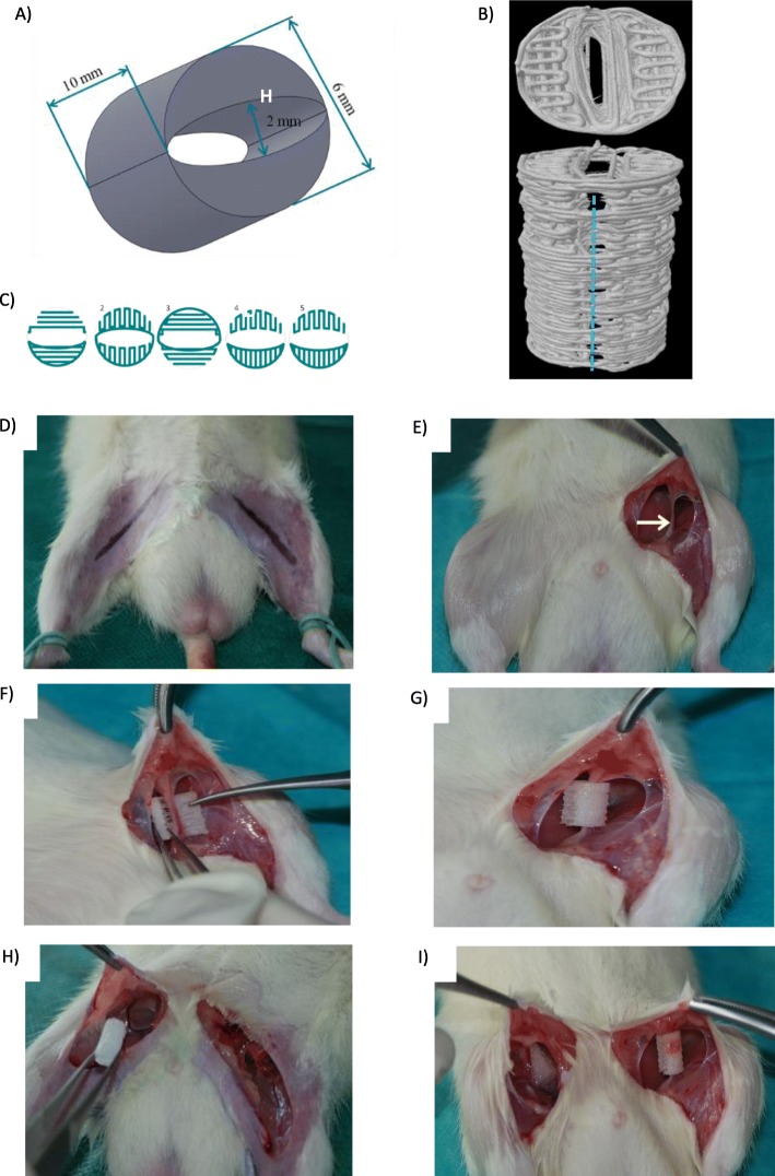
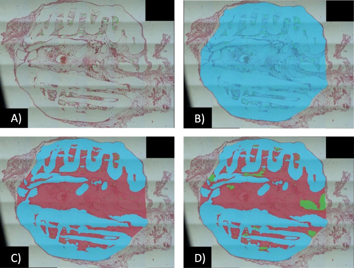
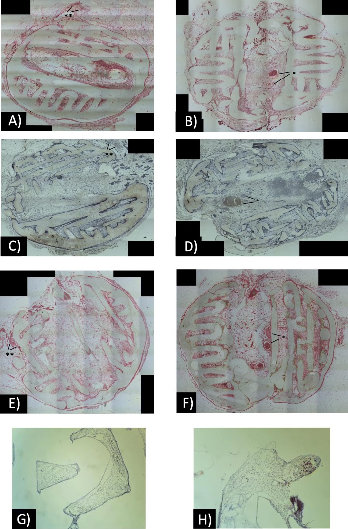
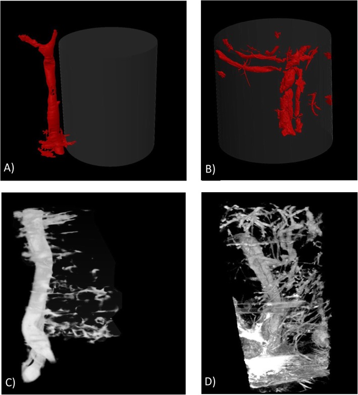
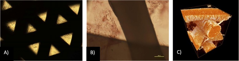
Methods: For prefabrication, functional blood vessels were introduced into the scaffold using a flow-through pedicle system. ASCs were isolated from rat fat deposits. Three-dimensional-printed cylindrical poly-ε-caprolactone scaffolds were fabricated by fused deposition modelling. Three groups, each containing six rats, were investigated by using non-seeded, ASC-seeded, and osteogenic induced ASC-seeded scaffolds. In each group, one rat was implanted with two scaffolds in the inguinal region. On the right side, a scaffold was implanted subcutaneously around the inferior epigastric vessels (classic prefabrication group). On the left side, the inferior epigastric vessels were placed inside the prefabricated scaffold in the flow-through pedicle system (flow-through prefabrication group). The vessel density and vascular architecture were examined histopathologically and by μCT imaging, respectively, at 2 months after implantation.

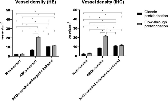
Results: The mean vessel densities were 10- and 5-fold higher in the ASC-seeded and osteogenic induced ASC-seeded scaffolds with flow-through prefabrication, respectively, than in the non-seeded classic prefabricated group (p < 0.001). μCT imaging revealed functional vessels within the scaffold.

Conclusion: ASC-seeded scaffolds with prefabrication showed significantly improved scaffold vasculogenesis and could be useful for application to tissue engineering products in the clinical settings.

Keywords: 3D printing; Flow-through pedicle; Prefabrication; Scaffold; Scaffold vascularization; Stem cells; Tissue engineering.


Conflict of interest statement

The authors declare that they have no competing interests.

Figures

Fig. 1
Fig. 1
Concept of the flow-through prefabrication method (a, b) and study design (c, d): a Vascular pedicle types: arteriovenous loop, arteriovenous bundle and flow-through pedicle. b New prefabrication method examined by the 3D reconstruction of μCT imaging showing the incision containing the scaffold from one side and its opening and the insertion of the flow-through type vascular pedicle into it. c Three groups of six rats each were used: a non-seeded group, an ASC-seeded group and an ASC-seeded osteogenic induced group. d Two methods of prefabrication were tested in each rat: classic prefabrication with the vascular pedicle outside the scaffold (vessel ingrowth only from outside) and flow-through prefabrication with the vascular pedicle inside the scaffold with a flow-through pedicle model (vessel ingrowth from outside and inside)
Fig. 2
Fig. 2
Scaffold design (a–c) and scaffold implantation (d–i): a Scaffold dimensions. b Place in the opening of the scaffold (blue line) used to insert the vascular pedicle inside (μCT 3D reconstruction model). c The fibre pattern was repeated every five layers during scaffold printing. d Incision markings. e Isolation of the vascular pedicle with the surrounding fascia. f Insertion of the vascular pedicle inside the scaffold. g Scaffold “closing” on the vascular pedicle. h Classic prefabrication group—insertion of the scaffold in the proximity of the vascular pedicle. i Correct placement of scaffolds: on the left side—vascular pedicle is inside the scaffold (flow-through prefabrication group); on the right side, vascular pedicle is outside but close to the scaffold (classic prefabrication group)
Fig. 3
Fig. 3
Vascular density analysis: a cross-sectional area with the counted number of vessels (N) (green crosses). b Specimen cross-sectional surface (S1). c Scaffold surface (blue area) (S2). d Artefact and empty area surface (green area) (S3)
Fig. 4
Fig. 4
Vessel density analysis was performed using both HE- (a, b) and CD31-stained(c, d) sections. The vascular pedicles were visible inside (*) or outside (**) in the flow-through (b, d) or classic prefabrication group, respectively. Bone formation assessed on von Kossa-stained (e, f) and SATB2-immmunostained (g, h) sections was not detected in any groups
Fig. 5
Fig. 5
Vessel density assessed on HE-stained (HE) and CD31-stained (IHC) sections. Notes: statistical analysis: * p < 0.001
Fig. 6
Fig. 6
Scaffold vascularity imaging: The classic (a) and flow-through prefabrication (b) groups were assessed through post-mortem and in vivo μCT imaging (c, d respectively). Small vessels with perfusion that sprouted from the main vascular pedicle were observed in the flow-through prefabrication group (b, d). In the classic prefabrication group, vessel ingrowth was noted only on the outer surface of the scaffold adjacent to the vascular pedicle (a, c)
Fig. 7
Fig. 7
In vitro studies on ASCs seeded PCL scaffolds: a, b Representative images of ASCs seeded into PCL after 21 days of culture. Alizarin red staining and mineralization assays of rat ASCs after 21 days: a control, non-differentiation culture of rat ASCs, b osteogenic differentiation. Seeding density of ASCs: 0.9 × 106/scaffold. Representative images are shown at × 10 magnification. Scale bars represent 100 μm. Images were obtained using an Olympus CKX41 microscope. c Typical μCT images of the PCL and scaffolds seeded with ASCs and incubated for 21 days. The images were reconstructed and analysed using the SkyScan NRecon and CtAn software. The 3D images were generated using SkyScan CTvol

References

    1. Jain RK, Au P, Tam J, Duda DG, Fukumura D. Engineering vascularized tissue. Nat Biotechnol. 2005;23:821–823. doi: 10.1038/nbt0705-821. - DOI - PubMed
    1. Carmeliet P, Jain RK. Angiogenesis in cancer and other diseases. Nature. 2000;407:249–257. doi: 10.1038/35025220. - DOI - PubMed
    1. Bai F, Wang Z, Lu J, Liu J, Chen G, Lv R, et al. The correlation between the internal structure and vascularization of controllable porous bioceramic materials in vivo: a quantitative study. Tissue Eng Part A. 2010;16:3791–3803. doi: 10.1089/ten.tea.2010.0148. - DOI - PubMed
    1. Nör JE, Peters MC, Christensen JB, Sutorik MM, Linn S, Khan MK, et al. Engineering and characterization of functional human microvessels in immunodeficient mice. Lab Investig. 2001;81:453–463. doi: 10.1038/labinvest.3780253. - DOI - PubMed
    1. Richardson TP, Peters MC, Ennett AB. Polymeric system for dual growth factor delivery. Nat Biotechnol. 2001;19:1029–1034. doi: 10.1038/nbt1101-1029. - DOI - PubMed

Publication types