Review
. 2020 Mar 6;295(10):3347-3361.
doi: 10.1074/jbc.REV119.010155. Epub 2020 Jan 23.

Uncovering the activities, biological roles, and regulation of bacterial cell wall hydrolases and tailoring enzymes

Affiliations
Review

Uncovering the activities, biological roles, and regulation of bacterial cell wall hydrolases and tailoring enzymes

Truc Do et al. J Biol Chem. .

Abstract

Bacteria account for 1000-fold more biomass than humans. They vary widely in shape and size. The morphological diversity of bacteria is due largely to the different peptidoglycan-based cell wall structures that encase bacterial cells. Although the basic structure of peptidoglycan is highly conserved, consisting of long glycan strands that are cross-linked by short peptide chains, the mature cell wall is chemically diverse. Peptidoglycan hydrolases and cell wall-tailoring enzymes that regulate glycan strand length, the degree of cross-linking, and the addition of other modifications to peptidoglycan are central in determining the final architecture of the bacterial cell wall. Historically, it has been difficult to biochemically characterize these enzymes that act on peptidoglycan because suitable peptidoglycan substrates were inaccessible. In this review, we discuss fundamental aspects of bacterial cell wall synthesis, describe the regulation and diverse biochemical and functional activities of peptidoglycan hydrolases, and highlight recently developed methods to make and label defined peptidoglycan substrates. We also review how access to these substrates has now enabled biochemical studies that deepen our understanding of how bacterial cell wall enzymes cooperate to build a mature cell wall. Such improved understanding is critical to the development of new antibiotics that disrupt cell wall biogenesis, a process essential to the survival of bacteria.

Keywords: Lipid II; LytR-CpsA-Psr ligases; O-acetyltransferase; acetyltransferase; antibiotic development; antibiotics; cell wall biochemistry; hydrolase; peptidoglycan; peptidoglycan hydrolase; peptidoglycan hydrolase regulators; teichoic acid.


Conflict of interest statement

The authors declare that they have no conflicts of interest with the contents of this article

Figures
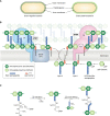
Figure 1.
Figure 1.
Overview of peptidoglycan assembly pathway. A, Gram-negative bacteria have an inner (IM) and outer (OM) cell membrane; a thin layer of peptidoglycan is sandwiched between the cell membranes. Gram-positive bacteria only have an inner cell membrane that is surrounded by a thick layer of peptidoglycan. B, Lipid II, the undecaprenyl pyrophosphate lipid-linked precursor for peptidoglycan synthesis, is assembled on the inner leaflet of the inner cell membrane. Once fully assembled, Lipid II is flipped to the outer leaflet of the inner cell membrane by a flippase. Peptidoglycan GTs polymerize Lipid II into glycan strands, which are cross-linked by transpeptidases (TP) into the existing matrix. There are two families of glycosyltransferases: the GT module of aPBPs and SEDS proteins. Class A PBPs are bifunctional, meaning they have polymerization and cross-linking activities. SEDS proteins cooperate with a partner bPBP, which only has transpeptidase activity. C, the active-site serine of PBP transpeptidases attacks the terminal d-Ala–d-Ala amide bond in a stem peptide (donor), forming an acyl-enzyme covalent intermediate and kicking out the terminal d-Ala. Resolution of the covalent intermediate occurs upon reaction with a nucleophilic amine from an incoming stem peptide (acceptor), producing a new peptide bond that links two glycan strands.
Figure 2.
Figure 2.
Cleavage sites of peptidoglycan hydrolases. Enzymes that hydrolyze peptidoglycan are broadly classified as glycosidases and peptidases, depending on where they cleave. Glycosidases (glucosaminidases, muramidases, and lytic transglycosylases) cleave within the glycan backbone. Peptidases (amidases, endopeptidases, ld-carboxypeptidases, and dd-carboxypeptidases) cleave peptide cross-links and within the stem peptides. The carets represent sites of hydrolysis.
Figure 3.
Figure 3.
Functions of peptidoglycan hydrolases. Bacterial predators weaponize hydrolases to degrade the peptidoglycan cell wall of their hosts, leading to host cell lysis. But hydrolases are more than just lysins. Bacteria harness endogenous hydrolases to support fundamental cellular processes. Peptidoglycan hydrolases play important roles in bacterial cell growth, differentiation, and the separation of daughter cells that have divided. They also tailor the peptidoglycan cell wall, controlling the length of glycan strands and the degree of cross-linking. Bacteria that recycle components of the cell wall use hydrolases to break the peptidoglycan matrix into smaller pieces that are transported back into the cell.
Figure 4.
Figure 4.
Direct regulation of peptidoglycan hydrolases. A (top), CwlV (PDB code 1JWQ) is an amidase that is active on its own. AmiB (PDB code 3NE8) and AmiC (PDB code 4BIN) are amidases that are directly activated by a regulator protein (69, 70). A helical domain, shown here in pink, sterically blocks the active site of AmiB and AmiC; displacement of this occluding helix activates the amidases. Bottom, a partial sequence alignment showing that the regulatory helix is present in AmiB and AmiC, but not CwlV. B, the E. coli amidases AmiA and AmiB are activated by direct interaction with EnvC, which itself is thought to be activated by FtsEX. In B. subtilis and S. pneumoniae, FtsEX is believed to regulate the hydrolases CwlO and PcsB, respectively. The ABC transporter FtsEX is presumed to harness ATP hydrolysis to adopt a conformation that is capable of activating the hydrolases.
Figure 5.
Figure 5.
Making and labeling peptidoglycan substrates to characterize cell wall enzymes. A, accumulation and extraction of Lipid II. Bacterial cultures are treated with an antibiotic that inhibits peptidoglycan synthesis to accumulate Lipid II in cells. The cells are spun down and resuspended in chloroform/methanol for the first extraction, which produces three layers. Lipid II is enriched in a thick interface fraction, whereas the majority of cellular phospholipids partition to the organic layer. A second extraction results in partitioning of Lipid II into the organic phase and UDP-MurNAc-pentapeptide, which is also present in the interface layer, into the aqueous phase. B, the two-step extraction allows isolation of Lipid II variants from the indicated species. C, uncross-linked glycan strands are synthesized from Lipid II using the monofunctional glycosyltransferase SgtB, which recognizes Lipid II variants with different stem peptides. To make cross-linked peptidoglycan, an appropriate bifunctional aPBP that recognizes the stem peptide is used. The aPBP polymerizes Lipid II and cross-links glycan strands. D, a chemical probe can be incorporated within the glycan backbone or stem peptide of peptidoglycan substrates to visualize and assess reaction of the substrates. i, one strategy to label the stem peptide of Lipid II or glycan strands makes use of the transpeptidase PBPX, which exchanges the terminal d-Ala in the stem peptide for a d-amino acid bearing a detectable tag. ii, another stem peptide labeling strategy selectively couples an amine-reactive probe to the side-chain primary amine at position 3 of the stem peptide. iii, a strategy to label the sugar backbone uses GalT to attach a [14C]galactose radiolabel at the nonreducing end of glycan strands. E, methods to digest peptidoglycan products for structural characterization by LC-MS. i, mutanolysin digestion and NaBH4 reduction of cross-linked peptidoglycan produce muropeptide species. ii, ColM treatment of uncross-linked glycan strands produces delipidated glycan diphosphate products.
Figure 6.
Figure 6.
Peptidoglycan hydrolases and tailoring enzymes have distinct substrate preferences. In S. aureus, the LCP wall teichoic acid ligases and the LytH-ActH amidase-activator complex are membrane-anchored proteins that only act on uncross-linked peptidoglycan substrates. The S. aureus O-acetyltransferase OatA is also a membrane-anchored protein and may act preferentially on uncross-linked peptidoglycan. By contrast, S. aureus Sle1 is an amidase that acts on cross-linked peptidoglycan. Another example of substrate selectivity is provided by lysozyme, which cleaves unacetylated, but not O-acetylated, backbones. Substrate selectivity can offer insights into the functions of these enzymes, as those enzymes that act at an earlier stage of peptidoglycan synthesis may show a preference for nascent peptidoglycan substrates.

References

    1. Schloss P. D., Girard R. A., Martin T., Edwards J., and Thrash J. C. (2016) Status of the archaeal and bacterial census: an update. MBio 7, e00201–16 10.1128/mBio.00201-16 - DOI - PMC - PubMed
    1. Bar-On Y. M., Phillips R., and Milo R. (2018) The biomass distribution on Earth. Proc. Natl. Acad. Sci. U.S.A. 115, 6506–6511 10.1073/pnas.1711842115 - DOI - PMC - PubMed
    1. Schulz H. N., and Jorgensen B. B. (2001) Big bacteria. Annu. Rev. Microbiol. 55, 105–137 10.1146/annurev.micro.55.1.105 - DOI - PubMed
    1. Young K. D. (2006) The selective value of bacterial shape. Microbiol. Mol. Biol. Rev. 70, 660–703 10.1128/MMBR.00001-06 - DOI - PMC - PubMed
    1. Young K. D. (2007) Bacterial morphology: why have different shapes? Curr. Opin. Microbiol. 10, 596–600 10.1016/j.mib.2007.09.009 - DOI - PMC - PubMed

Publication types

MeSH terms

LinkOut - more resources