. 2021 Feb 1;106(2):454-463.
doi: 10.3324/haematol.2019.220210.

Alternative activation of human macrophages enhances tissue factor expression and production of extracellular vesicles

Affiliations

Alternative activation of human macrophages enhances tissue factor expression and production of extracellular vesicles

Philipp J Hohensinner et al. Haematologica. .

Abstract

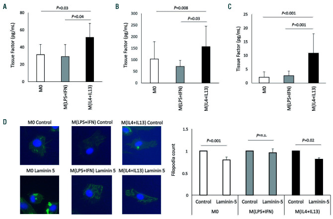
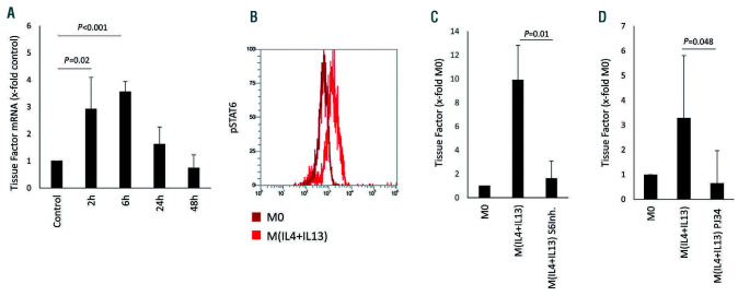
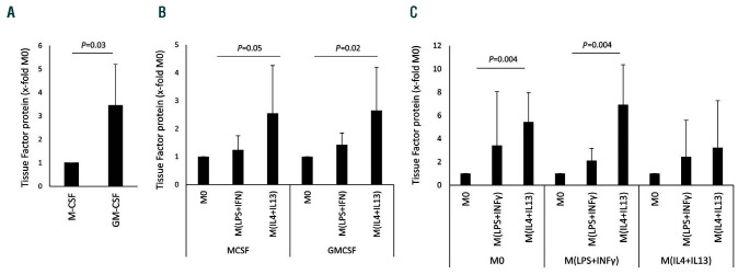
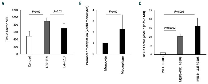
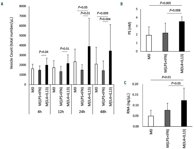
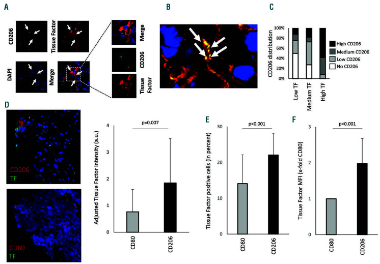
Macrophages are versatile cells that can be polarized by the tissue environment to fulfill required needs. Proinflammatory polarization is associated with increased tissue degradation and propagation of inflammation whereas alternative polarization within a Th2 cytokine environment is associated with wound healing and angiogenesis. To understand if polarization of macrophages can lead to a procoagulant macrophage subset we polarized human monocyte derived macrophages to a proinflammatory and an alternative activation state. Alternative polarization with interleukin-4 and IL-13 led to a macrophage phenotype characterized by increased tissue factor (TF) production and release and by an increase in extracellular vesicle production. In addition, also TF activity was enhanced in extracellular vesicles of alternatively polarized macrophages. This TF induction was dependent on signal transducer and activator of transcription-6 signaling and poly ADP ribose polymerase activity. In contrast to monocytes, human macrophages did not show increased tissue factor expression upon stimulation with lipopolysaccharide and interferon-γ. Previous polarization to either a proinflammatory or an alternative activation subset does not change the subsequent stimulation of TF. The inability of proinflammatory activated macrophages to respond to lipopolysaccharide and interferon-γ with an increase in TF production seems to be due to an increase in TF promoter methylation and was reversible when treating these macrophages with a demethylation agent. In conclusion, we provide evidence that proinflammatory polarization of macrophages does not lead to enhanced procoagulatory function, whereas alternative polarization of macrophages leads to an increased expression of TF and increased production of TF bearing extracellular vesicles by these cells suggesting a procoagulatory phenotype of alternatively polarized macrophages.


Figures

Figure 1.
Figure 1.
Tissue factor production after polarization of macrophages. (A) Tissue factor (TF) protein was determined on extracellular vesicles from supernatant (n=6) and (B) from lysed cells (n=7) using a specific enzyme-linked immunosorbent assay as indicated in the Methods section. (C) TF activity on extracellular vesicles from polarized macrophages was evaluated using an activity assay as indicated in the Methods section (n=13). Values are given in pg/mL and represent mean values ± standard deviation. (D) Capability of human polarized macrophages to form filopodia when migrating onto laminin-coated areas was evaluated by cytoskeletal staining (n=3). M0: unpolarized macrophages; M(LPS+IFN): classically activated polarized macrophages; M(IL-4+IL13): alternatively activated polarized macrophages; ns: not significant.
Figure 2.
Figure 2.
Tissue factor-specific mRNA induction by alternative polarization. (A) Tissue factor (TF) mRNA levels at the indicated time points in macrophages after polarization induced by interleukin (IL)-4 and IL-13 (n=3). (B) Phosphorylated STAT6 as determined in macrophages 30 min after IL-4+IL-13-induced polarization in comparison to that in unpolarized M0 macrophages using flow cytometry and specific antibodies as described in the Methods (n=3). A representative image is shown. (C) TF mRNA levels in macrophages 2 h after IL-4+IL-13-induced polarization in the presence and absence of a specific STAT6-inhibitor (S6Inh.) at 250 mM (n=6). (D) TF mRNA levels in macrophages 2 h after IL-4+IL-13-induced polarization in the presence and absence of the PARP-inhibitor PJ34 at 100 mM (n=6). TF mRNA in panels A, C and D was determined by quantitative polymerase chain reaction and GAPDH was used as a housekeeping gene as indicated in the Methods section. Values are given as fold changes compared to the respective unpolarized control (M0) and represent mean values ± standard deviation.
Figure 3.
Figure 3.
Tissue factor protein induction in different macrophage populations. (A) Tissue factor (TF) protein levels in supernatants from M0 macrophages generated via stimulation with macrophage colony-stimulation factor (MCSF) or granulocyte-macrophage colony-stimulating factor (GMCSF) (n=6). (B) TF protein in supernatants from macrophages that were generated either by stimulation with MCSF or GMCSF and polarized into M(LPS+IFN) and M(IL-4+IL-13) as indicated in the Methods. The respective M0 was used to determine the fold changes induced by the polarization conditions (n=6). (C) Macrophages were polarized for 48 h and afterwards repolarized for 24 h as indicated (n=5). TF protein levels were determined using a specific enzyme-linked immunosorbent assay as indicated in the Methods section. Values are given as fold changes compared to MCSF-differentiated macrophages in panel (A) or the respective unpolarized control (M0) in panels (B) and (C) and represent mean values ± standard deviation. M0: unpolarized macrophages; M(LPS+IFNγ): classically activated polarized macrophages; M(IL-4+IL13): alternatively activated polarized macrophages.
Figure 4.
Figure 4.
Epigenetic regulation of tissue factor. (A) Tissue factor (TF) expression on the surface of human monocytes cultured for 24 h in the presence of 100 ng/mL lipopolysaccharide (LPS) and 100 ng/mL interferon (IFN)-γ or 20 ng/mL interleukin (IL)-4 and 20 ng/mL IL-13 or without any addition (control) was analyzed by flow cytometry using a specific antibody as described in the Methods. Data are shown as mean fluorescence intensity (MFI) (n=4). (B) Methylation of the TF-promoter was analyzed by quantitative polymerase chain reaction as indicated in the Methods section. Macrophage values are given as fold increases compared to the monocyte value, which was set at 1. Monocytes and macrophages from the same individuals were compared (n=4). (C) TF protein was determined using a specific enzymelinked immunosorbent assay as indicated in the Methods section in the presence of the demethylating agent RG108 at a concentration of 5 mM during polarization. Values are given as fold changes compared to the respective unpolarized control (M0) (n=3) and represent mean values ± standard deviation. MFI: mean fluorescence intensity; M0: unpolarized macrophages; M(LPS+IFN): classically activated polarized macrophages; M(IL-4+IL13): alternatively activated polarized macrophages.
Figure 5.
Figure 5.
Influence of polarization conditions on extracellular vesicle production of human macrophages. (A) Numbers of extracellular vesicles in the supernatant of unpolarized macrophages and macrophages polarized in the presence of 100 ng/mL lipopolysaccharide (LPS) and 100 ng/mL interferon (IFN)-γ or 20 ng/mL interleukin (IL)-4 and 20 ng/mL IL-13 were determined by flow cytometry as indicated in the Methods section. Values are given as total vesicle count per mL: n=9 for 4 h and 12 h; n=12 for 24 h; and n=14 for 48 h. (B) Phosphatidylserine (PS) content of extracellular vesicles in the supernatant of M0 and macrophages polarized in the presence of 100 ng/mL LPS and 100 ng/mL IFN-γ or 20 ng/mL IL-4 and 20 ng/mL IL-13 for 48 h was determined using a specific enzyme-linked immunosorbent assay as indicated in the Methods. Values are given in nM PS (n=6). (C) Total extracellular vesicle-derived RNA was evaluated in the supernatant of M0 and macrophages polarized in the presence of 100 ng/mL LPS and 100 ng/mL IFN-γ or 20 ng/mL IL-4 and 20 ng/mL IL-13 for 48 h as indicated in the Methods section. Values are given in ng/mL RNA (n=5) and represent mean values ± standard deviation. M0: unpolarized macrophages; M(LPS+IFN): classically activated polarized macrophages; M(IL-4+IL13): alternatively activated polarized macrophages.
Figure 6.
Figure 6.
Staining of tissue factor-positive macrophages in sections of colon carcinoma. (A) Tissue factor (TF) (red) and CD206 (green) were stained in colon cancer tissue using specific antibodies as described in the Methods. CD206+ macrophages positive for TF are indicated with white arrows. The boxed area is shown in more detail. (B) White arrows indicate acellular regions that showed double staining for CD206 and TF (orange), which could represent extracellular vesicles derived from macrophages positive for CD206 and TF. (C) Distribution of CD206+ macrophages was evaluated and scored in areas with low, medium, and high TF density. (D) Human atherosclerotic plaque tissue was stained for TF (green) and either CD206 (red) alternatively activated macrophages or CD80 (red) for proinflammatory macrophages. Adjusted TF intensity to macrophage intensity demonstrated an increase in TF in CD206+ regions. Values are given as adjusted tissue factor intensity (arbitrary units) mean values ± standard deviation (SD) (n=16 patients). (E, F) Mouse macrophages from atherosclerotic plaques were isolated as indicated in the Online Supplement. Proinflammatory macrophages were less positive for TF and showed reduced mean fluorescence intensity compared to alternatively activated CD206 macrophages (n=11). Values represent mean values ± SD. DAPI: 4',6-diamidino-2-phenylindole; a.u.: arbitrary units.

References

    1. Glass CK. Genetic and genomic approaches to understanding macrophage identity and function. Arterioscler Thromb Vasc Biol. 2015;35(4):755-762. - PMC - PubMed
    1. Cybulsky MI, Cheong C, Robbins CS. Macrophages and dendritic cells: partners in atherogenesis. Circ Res. 2016;118(4):637-652. - PubMed
    1. Murray PJ, Wynn TA. Protective and pathogenic functions of macrophage subsets. Nat Rev Immunol. 2011;11(11):723-737. - PMC - PubMed
    1. Murray PJ. Macrophage polarization. Annu Rev Physiol. 2017;79:541-66. - PubMed
    1. Lech M, Anders HJ. Macrophages and fibrosis: how resident and infiltrating mononuclear phagocytes orchestrate all phases of tissue injury and repair. Biochim Biophys Acta. 2013;1832(7):989-997. - PubMed

Publication types