. 2020 Jan 23;10(1):1074.
doi: 10.1038/s41598-020-57745-w.

Restoring Wnt6 signaling ameliorates behavioral deficits in MeCP2 T158A mouse model of Rett syndrome

Affiliations

Restoring Wnt6 signaling ameliorates behavioral deficits in MeCP2 T158A mouse model of Rett syndrome

Wei-Lun Hsu et al. Sci Rep. .

Abstract

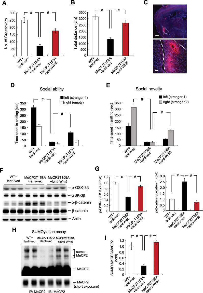
The methyl-CpG-binding protein 2 gene, MECP2, is an X chromosome-linked gene encoding the MeCP2 protein, and mutations of MECP2 cause Rett syndrome (RTT). Previous study has shown that re-expression of SUMO-modified MeCP2 in Mecp2-null neurons rescues synaptic and behavioral deficits in Mecp2 conditional knockout mice, whereas about 12-fold decrease in Wnt6 mRNA level was found in MeCP2K412R sumo-mutant mice. Here, we examined the role of Wnt6 in MeCP2 T158A mouse model of RTT. Results show that lentiviral delivery of Wnt6 to the amygdala ameliorates locomotor impairment and social behavioral deficits in these animals. MeCP2 T158A mice show decreased level of GSK-3β phosphorylation and increased level of β-catenin phosphorylation. They also show reduced level of MeCP2 SUMOylation. These alterations were also restored by lenti-Wnt6 transduction. Further, both BDNF and IGF-1 expressions are decreased in MeCP2 T158A mice. Overexpression of Wnt6 increases Bdnf and Igf-1 promoter activity in HEK293T cells in a dose-dependent manner. Lenti-Wnt6 transduction to the amygdala similarly increases the mRNA level and protein expression of BDNF and IGF-1 in MeCP2 T158A mice. Moreover, environmental enrichment (EE) similarly ameliorates the locomotor and social behavioral deficits in MeCP2 T158A mice. One of the mechanisms underlying EE is mediated through enhanced MeCP2 SUMOylation and increased Wnt6 expression in these animals by EE.


Conflict of interest statement

The authors declare no competing interests.

Figures

Figure 1
Figure 1
Overexpression of Wnt6 to the amygdala reduces locomotor impairment and social behavior deficits in MeCP2 T158A mutant mice. Animals were divided to three groups: WT mice received lenti-vector transduction, MeCP2 T158A mice received lenti-vector transduction and MeCP2 T158A mice received lenti-Wnt6 transduction to the amydgala. They were subjected to locomotor activity measure for 20 min 12 days later (n = 8 each group) and the (A) number of crossovers (F2,21 = 36.89, P < 0.001; q = 12.1, P < 0.001 comparing the WT + lenti-vector group with MeCP2 T158A + lenti-vector group and q = 7.03, P < 0.001 comparing the MeCP2 T158A + lenti-vector group with MeCP2 T158A + lenti-Wnt6 group) and (B) total distance travelled (F2,21 = 19.77, P < 0.001; q = 8.58, P < 0.001 comparing the WT + lenti-vector group with MeCP2 T158A + lenti-vector group and q = 6.31, P < 0.001 comparing the MeCP2 T158A + lenti-vector group with MeCP2 T158A + lenti-Wnt6 group) are shown. (C) Immunohistochemistry showing the location of lenti-mRFP-Wnt6 transduction and expression in the mouse amygdala. Scale bar equals 500 μm for the left panel and scale bar equals 100 μm for the right panel. The same animals were subjected to the (D) social ability test (for sniffing to stranger 1, F2,19 = 128.65, P < 0.001; q = 21.72, P < 0.001 comparing the WT + lenti-vector group with MeCP2 T158A + lenti-vector group and q = 5.59, P < 0.001 comparing the MeCP2 T158A + lenti-vector group with MeCP2 T158A + lenti-Wnt6 group) and (E) social novelty test (for sniffing to stranger 2, F2,19 = 107.48, P < 0.001; q = 20.28, P < 0.001 comparing the WT + lenti-vector group with MeCP2 T158A + lenti-vector group and q = 6.79, P < 0.001 comparing the MeCP2 T158A + lenti-vector group with MeCP2 T158A + lenti-Wnt6 group) one week later (n = 7 or 8 each group). (F) The amygdala tissue from these animals was dissected out and subjected to western blot analyses for the expression of phospho(p)Ser-9 GSK-3β, GSK-3β, phospho(p)Ser33/37/Thr41 β-catenin and β-catenin. A representative gel pattern is shown. (G) The quantified results are shown (n = 6 or 8 each group) (for p-GSK-3β over GSK-3β, F2,17 = 45.07, P < 0.001; q = 13.18, P < 0.001 comparing the WT + lenti-vector group with MeCP2 T158A + lenti-vector group and q = 9.23, P < 0.001 comparing the MeCP2 T158A + lenti-vector group with MeCP2 T158A + lenti-Wnt6 group) (for p-β-catenin over β-catenin, F2,17 = 27.4, P < 0.001; q = 8.69, P < 0.001 comparing the WT + lenti-vector group with MeCP2 T158A + lenti-vector group and q = 9.58, P < 0.001 comparing the MeCP2 T158A + lenti-vector group with MeCP2 T158A + lenti-Wnt6 group). (H) The same amygdala tissue lysates used above were also subjected to MeCP2 SUMOylation determination. A representative gel pattern is shown. (I) The quantified results are shown (n = 4 each group) (F2,9 = 26.25, P < 0.001; q = 8.05, P < 0.001 comparing the WT + lenti-vector group with MeCP2 T158A + lenti-vector group and q = 9.52, P < 0.001 comparing the MeCP2 T158A + lenti-vector group with MeCP2 T158A + lenti-Wnt6 group). Data are expressed as mean ± SEM. #P < 0.001.
Figure 2
Figure 2
Overexpression of Wnt6 increases the promoter activity and expression level of BDNF and IGF-1. (A) Bdnf promoter-luciferase construct containing the WRE element is shown. (B) Igf-1 promoter-luciferase construct containing the WRE element is shown. (C) Different amount of Flag-Wnt6 plasmid together with the pTALuc-Bdnf promoter construct (0.55 μg) and Renilla luciferase construct (0.05 μg) were transfected to HEK293T cells and Bdnf promoter activity was measured by luciferase assay (F3,16 = 257.03, P < 0.001). (D) Different amount of Flag-Wnt6 plasmid together with the pTALuc-Igf-1 promoter construct (0.55 μg) and Renilla luciferase construct (0.05 μg) were transfected to HEK293T cells and Igf-1 promoter activity was measured by luciferase assay (F3,16 = 183.53, P < 0.001). Western blot using anti-Flag antibody was adopted to confirm the transfection and expression of Flag-Wnt6 plasmids. Results are from five independent experiments. Lenti-vector was transducted to the amygdala of WT mice or MeCP2 T158A mice and lenti-Wnt6 was transducted to the amygdala of MeCP2 T158A mice. The amygdala tissue was dissected out and subjected to q-PCR analysis of (E) Bdnf mRNA level (F2,17 = 5.22, P < 0.05; q = 3.56, P < 0.05 comparing the WT + lenti-vector group with MeCP2 T158A + lenti-vector group and q = 4.32, P < 0.05 comparing the MeCP2 T158A + lenti-vector group with MeCP2 T158A + lenti-Wnt6 group) and Igf-1 mRNA level (F2,17 = 5.35, P < 0.05; q = 4.5, P < 0.05 comparing the WT + lenti-vector group with MeCP2 T158A + lenti-vector group and q = 3.31, P < 0.05 comparing the MeCP2 T158A + lenti-vector group with MeCP2 T158A + lenti-Wnt6 group). (F) V5-MeCP2WT, V5-MeCP2T158M or V5-MeCP2T158M + Flag-Wnt6 plasmids were transfected to the mouse amygdala and ChIP assay for CREB binding to the Bdnf and Igf-1 promoters are shown (n = 4 each group) (for CREB binding to the Bdnf promoter, F2,9 = 108.95, P < 0.001; q = 19.49, P < 0.001 comparing the WT + lenti-vector group with MeCP2 T158A + lenti-vector group and q = 16.21, P < 0.001 comparing the MeCP2 T158A + lenti-vector group with MeCP2 T158A + lenti-Wnt6 group) (for CREB binding to the Igf-1 promoter, F2,9 = 87.13, P < 0.001; q = 17.17, P < 0.001 comparing the WT + lenti-vector group with MeCP2 T158A + lenti-vector group and q = 14.92, P < 0.001 comparing the MeCP2 T158A + lenti-vector group with MeCP2 T158A + lenti-Wnt6 group). (G) The amygdala tissue from the same animals was also subjected to western blot determination of BDNF and IGF-1 protein expression. A representative gel pattern is shown. (H) The quantified results are shown (n = 6 or 8 each group) (for BDNF over actin, F2,17 = 73.66, P < 0.001; q = 9.56, P < 0.001 comparing the WT + lenti-vector group with MeCP2 T158A + lenti-vector group and q = 17.16, P < 0.001 comparing the MeCP2 T158A + lenti-vector group with MeCP2 T158A + lenti-Wnt6 group) (for IGF-1 over actin, F2,17 = 38.76, P < 0.001; q = 11.78, P < 0.001 comparing the WT + lenti-vector group with MeCP2 T158A + lenti-vector group and q = 9.71, P < 0.001 comparing the MeCP2 T158A + lenti-vector group with MeCP2 T158A + lenti-Wnt6 group). Data are expressed as mean ± SEM. *P < 0.05 and #P ≤ 0.001.
Figure 3
Figure 3
Environmental environment (EE) upregulates MeCP2 SUMOylation and Wnt6, BDNF as well as IGF-1 expression and alleviates behavioral deficits in MeCP2 T158A mice. (A) Animals were divided to three groups: WT mice housed in the home cage, MeCP2 T158A mice housed in the home cage and MeCP2 T158A mice subjected to EE. They were subjected to locomotor activity measure for 20 min at the end of EE (n = 8 each group) (F2,21 = 20.52, P < 0.001; q = 9.02, P < 0.001 comparing the WT group with MeCP2 T158A group and q = 3.81, P < 0.05 comparing the MeCP2 T158A group with MeCP2 T158A + EE group). The same animals were subjected to the (B) social ability test (for sniffing to stranger 1, F2,19 = 19.39, P < 0.001; q = 8.72, P < 0.001 comparing the WT group with MeCP2 T158A group and q = 3.47, P < 0.05 comparing the MeCP2 T158A group with MeCP2 T158A + EE group) and (C) social novelty test (for sniffing to stranger 2, F2,19 = 17.84, P < 0.001; q = 8.39, P < 0.001 comparing the WT group with MeCP2 T158A group and q = 3.5, P < 0.05 comparing the MeCP2 T158A group with MeCP2 T158A + EE group) one week later (n = 7 or 8 each group). (D) The amygdala tissue from these animals was dissected out and subjected to MeCP2 SUMOylation assay. The quantified result is shown at the lower panel (n = 6 or 8 each group) (F2,17 = 110.95, P < 0.001; q = 20.56, P < 0.001 comparing the WT group with MeCP2 T158A group and q = 14.87, P < 0.001 comparing the MeCP2 T158A group with MeCP2 T158A + EE group). (E) The same tissue lysate was also subjected to western blot determination of Wnt6, BDNF and IGF-1 expression. (F) The quantified results of Wnt6, BDNF and IGF-1 expression are shown (n = 6 or 8 each group) (for Wnt6, F2,17 = 87.93, P < 0.001; q = 15.79, P < 0.001 comparing the WT group with MeCP2 T158A group and q = 16.99, P < 0.001 comparing the MeCP2 T158A group with MeCP2 T158A + EE group) (for BDNF, F2,17 = 36.94, P < 0.001; q = 11.52, P < 0.001 comparing the WT group with MeCP2 T158A group and q = 9.44, P < 0.001 comparing the MeCP2 T158A group with MeCP2 T158A + EE group) (for IGF-1, F2,17 = 21.17, P < 0.001; q = 7.87, P < 0.001 comparing the WT group with MeCP2 T158A group and q = 8.24, P < 0.001 comparing the MeCP2 T158A group with MeCP2 T158A + EE group). (G) Mice received PBS or NMDA (6 mM) injection to their amygdala and Wnt6 expression was determined by western blot 1 h later (n = 6 each group) (t1,10 = 20.52, P < 0.001). Data are expressed as mean ± SEM. *P < 0.05 and #P < 0.001.
Figure 4
Figure 4
Schematic diagram showing that Wnt6 signaling is impaired in MeCP2 T158A mice and Wnt6 expression rescues the locomotor activity impairment and social behavior deficits in MeCP2 T158A mice. Further, Wnt6 expression is regulated by environmental enrichment. EE: environmental enrichment.

Similar articles

Cited by

References

    1. Amir RE, et al. Rett syndrome is caused by mutations in X-linked MECP2, encoding methyl-CpG-binding protein 2. Nature genetics. 1999;23:185–188. doi: 10.1038/13810. - DOI - PubMed
    1. Nan X, et al. Transcriptional repression by the methyl-CpG-binding protein MeCP2 involves a histone deacetylase complex. Nature. 1998;393:386–389. doi: 10.1038/30764. - DOI - PubMed
    1. Chahrour M, et al. MeCP2, a key contributor to neurological diseases, activates and represses transcription. Science. 2008;320:1224–1229. doi: 10.1126/science.1153252. - DOI - PMC - PubMed
    1. Guy J, Cheval H, Selfridge J, Bird A. The role of MeCP2 in the brain. Annual review of cell and developmental biology. 2011;27:631–652. doi: 10.1146/annurev-cellbio-092910-154121. - DOI - PubMed
    1. Moretti P, Zoghbi HY. MeCP2 dysfunction in Rett syndrome and related disorders. Current opinion in genetics & development. 2006;16:276–281. doi: 10.1016/j.gde.2006.04.009. - DOI - PubMed

Publication types

MeSH terms