. 2020 Apr;14(4):1015-1029.
doi: 10.1038/s41396-020-0587-5. Epub 2020 Jan 23.

Mycelial network-mediated rhizobial dispersal enhances legume nodulation

Affiliations

Mycelial network-mediated rhizobial dispersal enhances legume nodulation

Wei Zhang et al. ISME J. 2020 Apr.

Abstract

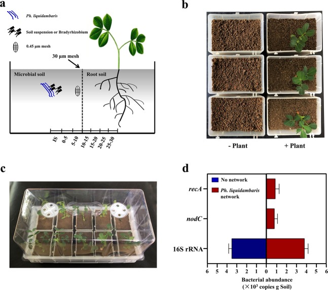
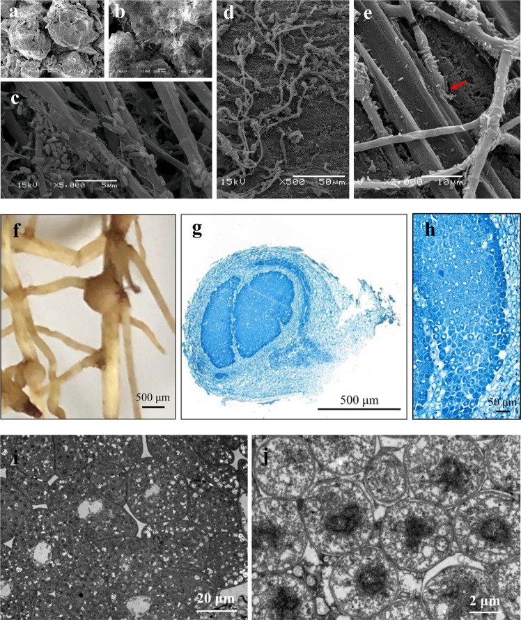
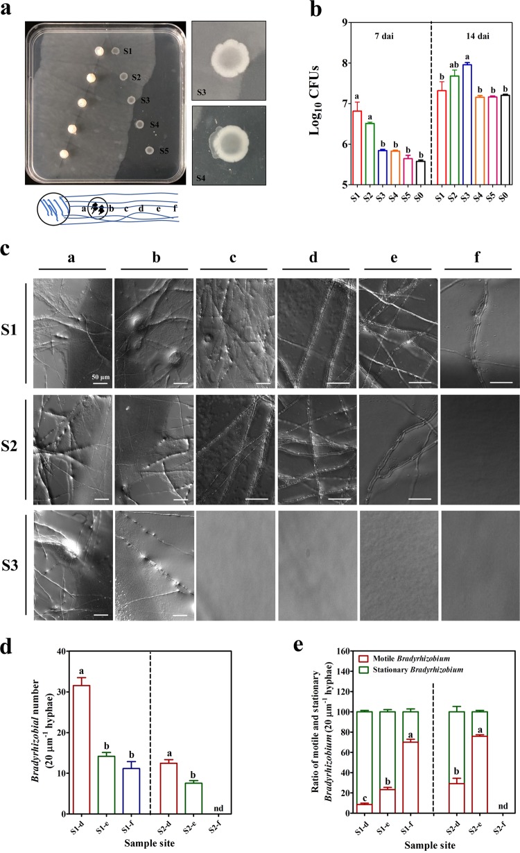
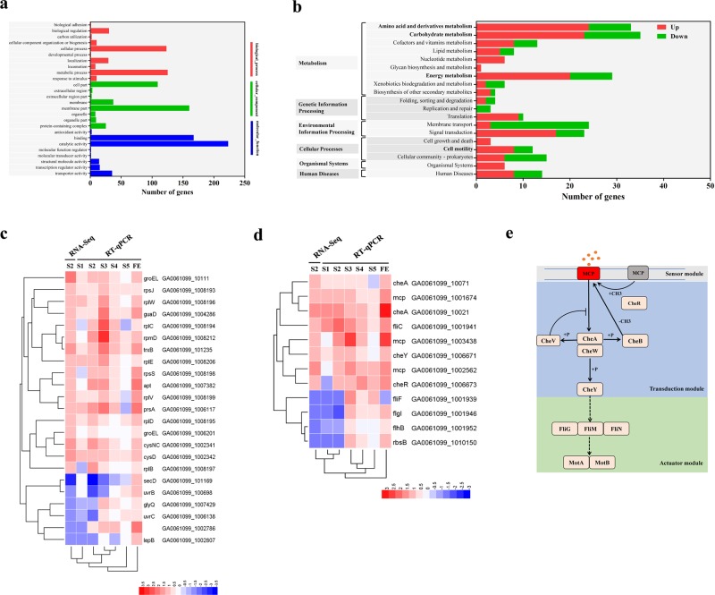
The access of rhizobia to legume host is a prerequisite for nodulation. Rhizobia are poorly motile in soil, while filamentous fungi are known to grow extensively across soil pores. Since root exudates-driven bacterial chemotaxis cannot explain rhizobial long-distance dispersal, mycelia could constitute ideal dispersal networks to help rhizobial enrichment in the legume rhizosphere from bulk soil. Thus, we hypothesized that mycelia networks act as vectors that enable contact between rhizobia and legume and influence subsequent nodulation. By developing a soil microcosm system, we found that a facultatively biotrophic fungus, Phomopsis liquidambaris, helps rhizobial migration from bulk soil to the peanut (Arachis hypogaea) rhizosphere and, hence, triggers peanut-rhizobium nodulation but not seen in the absence of mycelia. Assays of dispersal modes suggested that cell proliferation and motility mediated rhizobial dispersal along mycelia, and fungal exudates might contribute to this process. Furthermore, transcriptomic analysis indicated that genes associated with the cell division, chemosensory system, flagellum biosynthesis, and motility were regulated by Ph. liquidambaris, thus accounting for the detected rhizobial dispersal along hyphae. Our results indicate that rhizobia use mycelia as dispersal networks that migrate to legume rhizosphere and trigger nodulation. This work highlights the importance of mycelial network-based bacterial dispersal in legume-rhizobium symbiosis.


Conflict of interest statement

The authors declare that they have no conflict of interest.

Figures

Fig. 1
Fig. 1. Ph. liquidambaris transfers soil rhizobia to the rhizosphere of peanut.
a, b A soil microcosm system containing microbial and root compartments was established to determine whether Ph. liquidambaris networks transfer rhizobia to the rhizosphere of peanut. The microbial and root compartments were separated by a sterile 30 μm mesh. Roots were confined to the root soil, but fungi and rhizobia in the microbial soil were able to cross the mesh and enter root soil. The treatments with peanut cultivation contained individual seedling planted in root compartment. Sterile 0.45 μm mesh in soil microcosm was used to observe Ph. liquidambarisBradyrhizobium interaction. c Microcosms were placed in transparent boxes with closed lids to minimize water evaporation and avoid microbial contamination. d 16S rRNA, nodC, and recA copies in the rhizosphere soil of peanut. At 15 dai, rhizosphere soil of peanut was collected to determine the copy numbers of 16S rRNA, nodC, and recA when the microbial compartment was inoculated with soil suspension with or without Ph. liquidambaris. Data and error bars are the mean ± SE (n = 4). CFUs colony-forming units; dai days after inoculation; IS inoculation site.
Fig. 2
Fig. 2. Ph. liquidambaris facilitates Bradyrhizobial dispersal in soil conditions.
a Bradyrhizobial number was obtained by CFU determination from the soil at various collection distances at 3, 6, and 9 dai when the microbial compartment was inoculated with Bradyrhizobium with or without Ph. liquidambaris and the root compartment was planted with or without peanut. b Bradyrhizobial number was obtained by qPCR of nodC from the soil at various collection distances at 3, 6, and 9 dai when the microbial compartment was inoculated with Bradyrhizobium with or without Ph. liquidambaris and the root compartment was planted with or without peanut. Data and error bars are the mean ± SE (n = 4). CFU colony-forming units; dai days after inoculation.
Fig. 3
Fig. 3. Ph. liquidambaris facilitates Bradyrhizobial dispersal, promotes Bradyrhizobial root infection, and triggers peanut–Bradyrhizobium interaction.
a SEM image showing no fungal network in soil pores of microcosm without Ph. liquidambaris inoculation. b SEM image showing fungal networks formed by Ph. liquidambaris in soil pores of microcosm with Ph. liquidambaris inoculation. c SEM image showing the attachment of Bradyrhizobium on Ph. liquidambaris hyphae. Bradyrhizobium was attached on the hyphal surface and formed fiber-like biofilms in soil. At 9 dai, the meshes (0.45 μm) were sampled for scanning electron microscopy to observe Ph. liquidambarisBradyrhizobium interaction in soil environment. d SEM image showing Ph. liquidambarisBradyrhizobium interaction on the root surface. e SEM image showing the root invasion of hyphae-attached Bradyrhizobium through crack sites (red arrow). At 9 dai, peanut roots were sampled for scanning electron microscopy to observe Ph. liquidambarisBradyrhizobium interaction on root surface. f Nodules on the lateral root of peanut in the presence of Ph. liquidambaris and Bradyrhizobium. No visible nodule was observed on the root in the absence of Ph. liquidambaris. g Toluidine blue-stained transverse section of nodules. h Higher-magnification images from g. The infected cells were well organized in the N2-fixation zone. i, j TEM images showing the symbiosomes and bacteroides. The bacteroides were well organized in symbiosomes. At 45 dai, root nodules were collected and prepared for transmission electron microscopy to observe nodule ultrastructure. The experiments were carried out with three individual replicates, and representative micrographs are shown. SEM scanning electron microscope; TEM transmission electron microscope; dai days after inoculation.
Fig. 4
Fig. 4. Ph. liquidambaris networks help Bradyrhizobial dispersal.
a Plate with a sterile polystyrene ring with Bradyrhizobium inoculation alone. b Plate with a sterile polystyrene ring with Ph. liquidambaris and Bradyrhizobium co-inoculation. A–C sites refer to sampling sites for scanning electron microscopy. c, d SEM images showing Bradyrhizobium attached to and dispersed from mycelial networks of Ph. liquidambaris at A site. e, f SEM images showing Bradyrhizobium attached to and dispersal from mycelial networks at B site. g, h SEM images showing Bradyrhizobium attached to and dispersal from mycelial networks at C site. d, f, h Higher-magnification images from c, e, and g. The experiments were carried out with three individual replicates, and representative micrographs are shown. SEM scanning electron microscope.
Fig. 5
Fig. 5. Analysis of Bradyrhizobial dispersal on mycelial networks of Ph. liquidambaris at 14 dai.
a Ph. liquidambaris and Bradyrhizobium were inoculated on a square plate in a diagonal row. At 14 dai, Ph. liquidambaris began to interact with Bradyrhizobium at S3. At S4, Bradyrhizobium was attracted by Ph. liquidambaris before physical contact. The sketch refers to sampling sites of square plate above for microscopic observation. The big circle represents fungal plug and the small circle represents Bradyrhizobial colony. a–f in the sketch represent the sampling sites. a, b in the sketch represent the sampling sites at left and right of Bradyrhizobial colony. b Effects of Ph. liquidambaris on Bradyrhizobial growth at S0–S5 at 7 and 14 dai. Bradyrhizobial number was determined by CFU. Data and error bars are the mean ± SE (n = 3) and different letters indicate significant differences among different sites at the same dai (one-way analysis of variance with Tukey’s test, P < 0.05). c Different stages of Ph. liquidambarisBradyrhizobial interaction at 14 dai showing Bradyrhizobial dispersal along mycelial networks. Samples were collected from a and directly observed with a Zeiss Axio Imager A1 microscope. Bars, 50 μm. d Average number of Bradyrhizobium on hyphae of different sampling site of c. e Average ratio of motile and stationary Bradyrhizobium on hyphae of different sampling site of c. The number of total, motile, and stationary Bradyrhizobium on 20 μm hyphae was counted from images and time-lapse videos, respectively. The experiments were performed with three individual replicates and each replicate contained 15 images or time-lapse videos over 60 s. Data and error bars are the mean ± SE (n = 3) and different letters indicate significant differences among different sites (one-way analysis of variance with Tukey’s test, P < 0.05). dai days after inoculation.
Fig. 6
Fig. 6. Effects of Ph. liquidambaris exudates on Bradyrhizobial growth, biofilm formation, and chemotaxis.
a Ph. liquidambaris exudates increased Bradyrhizobial growth. The OD590 values were recorded every 4 h for 78 h. b Ph. liquidambaris exudates increased Bradyrhizobial biofilm formation. c Chemotactic response of Bradyrhizobium toward Ph. liquidambaris exudates. The chemotactic response of Bradyrhizobium toward Ph. liquidambaris exudates was evaluated by capillary assay. Data and error bars are the mean ± SE (n = 3) and asterisks indicate significant differences between control and fungal exudates treatments (Student’s t test, *P < 0.05, **P < 0.01, ***P < 0.001). CFU colony-forming units.
Fig. 7
Fig. 7. RNA-seq reveal the potential genetic mechanisms of rhizobial dispersal on Ph. liquidambaris networks.
a Number of differentially expressed genes of GO categories related to biological process, cellular component, and molecular function in Bradyrhizobium inoculation alone (B) vs Bradyrhizobium and Ph. liquidambaris co-inoculation (E + B). b KEGG analysis of differentially expressed genes in B vs E + B. c Expression of Bradyrhizobial differentially expressed genes associated with bacterial division at S0-S5 at 14 dai by RNA-seq and qRT-PCR. The responses of these differentially expressed genes to fungal exudates were also included. d Expression of Bradyrhizobial differentially expressed genes associated with bacterial chemotaxis, flagellar biosynthesis, and motility at S0–S5 at 14 dai by RNA-seq and qRT-PCR. The responses of these differentially expressed genes to fungal exudates were also included. e Bacterial swimming chemotaxis pathway, which includes sensor, transduction, and actuator modules. dai days after inoculation; FE fungal exudates.

Similar articles

Cited by

References

    1. Zipfel C, Oldroyd GED. Plant signalling in symbiosis and immunity. Nature. 2017;543:328–36. - PubMed
    1. Mus F, Crook MB, Garcia K, Costas AG, Geddes BA, Kouri ED, et al. Symbiotic nitrogen fixation and the challenges to its extension to nonlegumes. Appl Environ Microbiol. 2016;82:3698–710. - PMC - PubMed
    1. Burchill W, James EK, Li D, Lanigan GJ, Williams M, Iannetta PPM, et al. Comparisons of biological nitrogen fixation in association with white clover (Trifolium repens L.) under four fertilizer nitrogen inputs as measured using two 15N techniques. Plant Soil. 2014;385:287–302.
    1. López-García SL, Perticari A, Piccinetti C, Ventimiglia L, Arias N, De Battista JJ, et al. In-furrow inoculation and selection for higher motility enhances the efficacy of Bradyrhizobium japonicum nodulation. Agron J. 2009;101:357–63.
    1. Vicario JC, Dardanelli MS, Giordano W. Swimming and swarming motility properties of peanut-nodulating rhizobia. FEMS Microbiol Lett. 2015;362:1–6. - PubMed

Publication types