Comparative Study
. 2020 Jan 23;10(1):1039.
doi: 10.1038/s41598-020-57497-7.

A comparison of methods for the isolation and separation of extracellular vesicles from protein and lipid particles in human serum

Affiliations
Comparative Study

A comparison of methods for the isolation and separation of extracellular vesicles from protein and lipid particles in human serum

K Brennan et al. Sci Rep. .

Abstract

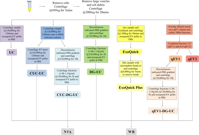
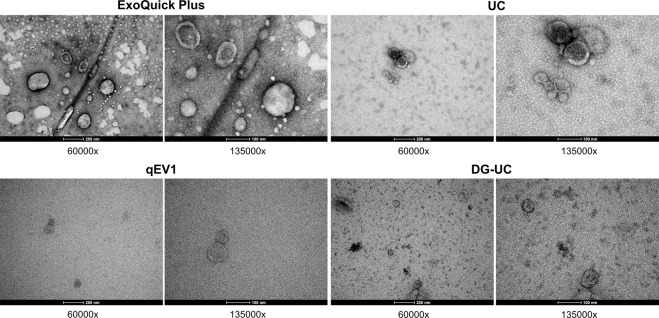
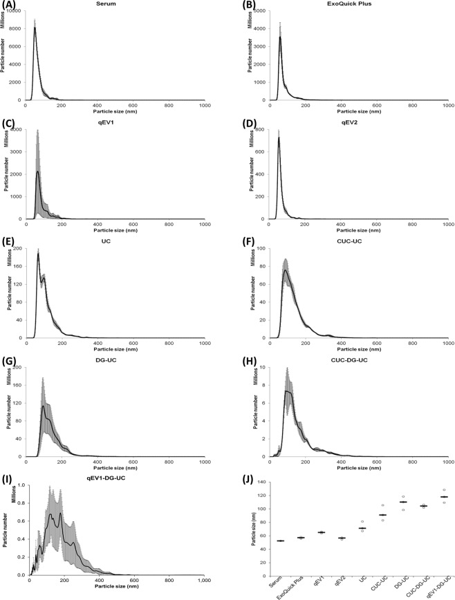
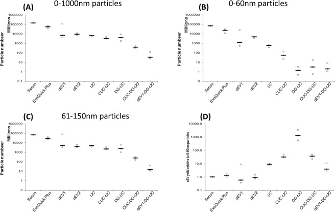
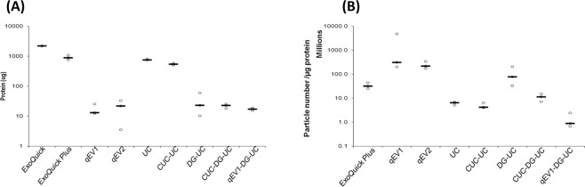
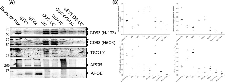
Extracellular vesicles (EVs) are nano-sized vesicles containing nucleic acid and protein cargo that are released from a multitude of cell types and have gained significant interest as potential diagnostic biomarkers. Human serum is a rich source of readily accessible EVs; however, the separation of EVs from serum proteins and non-EV lipid particles represents a considerable challenge. In this study, we compared the most commonly used isolation techniques, either alone or in combination, for the isolation of EVs from 200 µl of human serum and their separation from non-EV protein and lipid particles present in serum. The size and yield of particles isolated by each method was determined by nanoparticle tracking analysis, with the variation in particle size distribution being used to determine the relative impact of lipoproteins and protein aggregates on the isolated EV population. Purification of EVs from soluble protein was determined by calculating the ratio of EV particle count to protein concentration. Finally, lipoprotein particles co-isolated with EVs was determined by Western blot analysis of lipoprotein markers APOB and APOE. Overall, this study reveals that the choice of EV isolation procedure significantly impacts EV yield from human serum, together with the presence of lipoprotein and protein contaminants.


Conflict of interest statement

The authors declare no competing interests.

Figures

Figure 1
Figure 1
Schematic summary of EV isolation and downstream analyses. EVs were isolated from human serum using five different methods alone or in combination and characterized by Western blot (WB), nanoparticle tracking analysis (NTA).
Figure 2
Figure 2
Transmission electron microscopic (TEM) visualization of the EVs isolated from pooled human serum. Representative electron microscopy images of EVs isolated by ExoQuick Plus, ultracentrifugation (UC), size exclusion chromatography (qEV1), or density gradient ultracentrifugation (DG-UC), from pooled human serum. 60k and 135k magnifications are shown.
Figure 3
Figure 3
Nanosight analysis of particle size distribution of the EVs isolated from pooled human serum. Nanoparticle tracking analysis (NTA) of the total vesicles isolated from 200 µl of serum by each method alone or in combination. (AI) Line graphs corresponding to average number and size of particles isolated by each method, calculated from the mean of 15 videos per isolation.) (J) Dot plots representing the variation in the modal size of the vesicle isolated by each method. Black lines represent medians. N = 3 isolations.
Figure 4
Figure 4
Nanosight analysis of particle number of the EVs isolated from pooled human serum. Nanoparticle tracking analysis (NTA) of the total vesicles isolated from 200 µl of serum by each method alone or in combination. Dot plots corresponding to average numbers of vesicles between (A) 0–1000 nm, (B) 0–60 nm, and (C) 61–150 nm isolated by each method, calculated from the mean of 15 videos per isolation. (D) Dot plots corresponding to sEV (61–150 nm vesicle) number/ 0–60 nm vesicle number isolated by each method. Black lines represent medians. N = 3 isolations.
Figure 5
Figure 5
Serum protein co-isolated with EVs isolated from pooled human serum. (A) Dot plots representing the total amount of protein present in the EV isolations by each method alone or in combination was determined by BCA assay. N = 3 isolations (B) Dot plots corresponding to 61–150 nm vesicle number/µg of protein isolated by each method. Vesicle number was determined by nanoparticle tracking analysis (NTA) of 15 videos per isolation. Black lines represent medians. N = 3 isolations.
Figure 6
Figure 6
Western blot analysis of EV markers, CD63 and TSG101, and lipoprotein markers, APOB, and APOE. (A) 4 µg of vesicular protein isolated from 200 µl human serum using each method alone or in combination were separated by SDS–PAGE and immunoblotted with anti-human CD63 (H-193 and H5C6), TSG101, APOE and APOB antibodies. Representative images are shown. (B) Graph: Densitometric quantitation of the distribution of CD63, TSG101, APOE and APOB protein expression in each method. Black lines represent medians. N = 3 isolations. Arbitrary Units (AU) = (signal intensity method)/Sum (signal intensity method1–8).

References

    1. Harding C, Heuser J, Stahl P. Receptor-mediated endocytosis of transferrin and recycling of the transferrin receptor in rat reticulocytes. J. Cell. Biol. 1983;97:329–339. doi: 10.1083/jcb.97.2.329. - DOI - PMC - PubMed
    1. Rieu S, Geminard C, Rabesandratana H, Sainte-Marie J, Vidal M. Exosomes released during reticulocyte maturation bind to fibronectin via integrin alpha4beta1. Eur. J. Biochem. 2000;267:583–590. doi: 10.1046/j.1432-1327.2000.01036.x. - DOI - PubMed
    1. Pan BT, Teng K, Wu C, Adam M, Johnstone RM. Electron microscopic evidence for externalization of the transferrin receptor in vesicular form in sheep reticulocytes. J. Cell Biol. 1985;101:942–948. doi: 10.1083/jcb.101.3.942. - DOI - PMC - PubMed
    1. Thery C, Ostrowski M, Segura E. Membrane vesicles as conveyors of immune responses. Nat. Rev. Immunol. 2009;9:581–593. doi: 10.1038/nri2567. - DOI - PubMed
    1. Valadi H, et al. Exosome-mediated transfer of mRNAs and microRNAs is a novel mechanism of genetic exchange between cells. Nat. Cell Biol. 2007;9:654–659. doi: 10.1038/ncb1596. - DOI - PubMed

Publication types

MeSH terms