. 2020 Jan 23;3(1):41.
doi: 10.1038/s42003-020-0761-3.

Development of chimeric peptides to facilitate the neutralisation of lipopolysaccharides during bactericidal targeting of multidrug-resistant Escherichia coli

Affiliations

Development of chimeric peptides to facilitate the neutralisation of lipopolysaccharides during bactericidal targeting of multidrug-resistant Escherichia coli

Zhenlong Wang et al. Commun Biol. .

Abstract

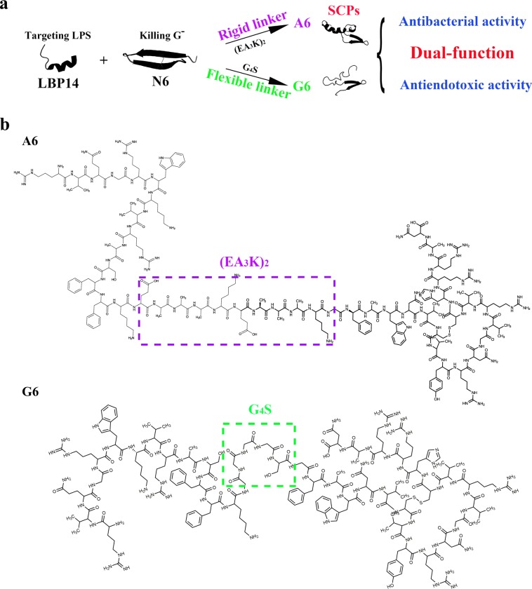
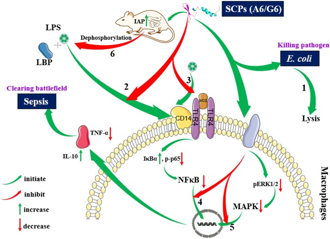
Pathogenic Escherichia coli can cause fatal diarrheal diseases in both animals and humans. However, no antibiotics or antimicrobial peptides (AMPs) can adequately kill resistant bacteria and clear bacterial endotoxin, lipopolysaccharide (LPS) which leads to inflammation and sepsis. Here, the LPS-targeted smart chimeric peptides (SCPs)-A6 and G6 are generated by connecting LPS-targeting peptide-LBP14 and killing domain-N6 via different linkers. Rigid and flexible linkers retain the independent biological activities from each component. SCPs-A6 and G6 exert low toxicity and no bacterial resistance, and they more rapidly kill multiple-drug-resistant E. coli and more effectively neutralize LPS toxicity than N6 alone. The SCPs can enhance mouse survival more effectively than N6 or polymyxin B and alleviate lung injuries by blocking mitogen-activated protein kinase and nuclear factor kappa-B p65 activation. These findings uniquely show that SCPs-A6 and G6 may be promising dual-function candidates as improved antibacterial and anti-endotoxin agents to treat bacterial infection and sepsis.


Conflict of interest statement

The authors declare no competing interests.

Figures

Fig. 1
Fig. 1. Design and structure scheme of SCPs-A6 and G6.
a Design schemes of SCPs by connecting LBP14 (a targeting domain) and N6 (a killing domain) via (EA3K)2/G4S linkers. b Structure schemes of A6 and G6.
Fig. 2
Fig. 2. Time-killing curves, stability, and resistance of SCPs-A6 and G6 against MDR E. coli CVCC195 and their toxicity toward eukaryotic cells.
a The time-killing curves of A6, G6 and N6 against E. coli CVCC195 in vitro. Control: PBS. PMB: polymyxin B. be Effects of temperature (b), pH (c), enzyme (d), serum (e) on the antibacterial activity of A6, G6, and N6 against E. coli CVCC195. f The peptide remaining in serum. g Resistance of A6 and G6. h The hemolysis of A6 and G6 against fresh mouse red blood cells. 0.1% Triton X-100 served as the positive control (100% hemolysis). i The cytotoxicity of A6 and G6 against RAW 264.7 monocytes. Results indicate means with SD (n = 3 independent experiments). Different lower case letters indicate a difference between two groups (p < 0.05). Source data for ad can be found in Supplementary Data.
Fig. 3
Fig. 3. Effects of SCPs-A6 and G6 on the cell morphologies and ultrastructures of MDR E. coli CVCC195.
Bacteria in mid-logarithmic growth were treated with peptides or antibiotic at 4 × MIC for 2 h (n = 3 independent experiments). a SEM images. The scalebar represents 2 and 1 μm. b TEM images. Both N6 and PMB were used as controls. Red arrows indicated typical disruptions, which was caused by peptides (filamentous substances, disappearance of membranes, leakage of contents and ghosts) or PMB (protrusions). The scalebar represents 5, 1, and 0.2 μm.
Fig. 4
Fig. 4. Interaction between SCPs-A6 or G6 and LPS.
a Dissociation of LPS aggregates. FITC-labeled LPS micells were incubated with A6, G6, N6, and PMB, and the fluorescent intensity was detected in a microplate reader. Results indicate means with SD (n = 3 independent experiments). b CD spectra for A6, G6, or N6 with or without E. coli LPS (0.2 mg per ml). c Binding affinity of LBP14, A6, G6, and N6 to LPS. Ampicillin (AMP) and PMB were used as negative and positive controls, respectively. Results indicate means with SD (n = 3 independent experiments). Different lower case letters indicate a difference between two groups (p < 0.05). Source data for ac can be found in Supplementary Data 1. d, e NMR analysis of A6 (d) and G6 (e) binding to LPS. Part of the NOESY spectra for A6 and G6 showed an NOE enhancement upon addition of LPS to the peptide solvent (black: free peptide; red: peptide with LPS). Some of the tr-NOEs for the targeting domain (residues 1–14 in A6 and G6) showed that intra-residue and/or sequential NOEs were labeled beside the peaks.
Fig. 5
Fig. 5. Effects of SCPs-A6 and G6 on LPS binding to LBP and macrophages.
a Effects of A6 and G6 on the binding of LPS and LBP. The 96-well microtiter plates were coated with 4 ng per ml LPS overnight at 4 °C, washed three times with PBST, and blocked with PBS for 2 h. After washing again, the plates were incubated with different concentrations of A6 and G6 at 37 °C for 1 h. After the addition of primary and secondary antibodies, the absorbance was measured at OD450 nm. N6 and PMB were used as controls. Results indicate means with SD (n = 3 independent experiments). Different lower case letters indicate a difference between two groups (p < 0.05). b Location of A6 and G6 in RAW 264.7 macrophages. Macrophages were preincubated with FITC-labeled LPS (100 μg per ml), washed, treated with rhodamine-labeled A6 and G6 (0.1 μM), and analyzed by CLSM. The scalebar indicates 7.5 and 10 μM. c Effects of A6 and G6 on the binding of LPS to RAW 264.7 cells. Cells were cultured overnight and incubated for 30 min at 37 °C with FITC-labeled LPS (100 μg per ml), followed by washing with PBS. Both A6 and G6 (0.1 μM) were added into cells, incubated for 1 h and analyzed by flow cytometry. N6 and PMB were used as controls. d Biodistribution of A6 and G6 in the healthy nude mice. The nude mice were injected intraperitoneally with 5 mg per kg FITC-labeled A6, G6, N6 or free FITC, and fluorescence (ventral) was observed at 0.5, 1, 2, 4, 8, 24, 48, and 72 h, respectively. The mice from left to right were free FITC, FITC-labeled N6, FITC-labeled A6, FITC-labeled G6 and the blank control, respectively. Source data for a can be found in Supplementary Data 1.
Fig. 6
Fig. 6. Efficacy of SCPs-A6 and G6 in mice challenged with MDR E. coli and LPS.
a Survival of mice. Mice were intraperitoneally injected with E. coli (5 × 108 CFU) or LPS (13.5 mg per kg of body weight), followed by injection with A6 and G6 at 0.5 and 8 h, respectively. N6 and PMB were used as controls. The mouse survival was recorded for 7 d (n = 5 independent mice). b Effects of A6 and G6 on the cytokines and IAP levels. The results are given as the mean ± SD (n = 3 independent experiments). Different lower case letters indicate a difference between two groups (p < 0.05). c Effects of A6 and G6 on lung injuries induced by LPS. Mice were injected intraperitoneally with LPS and were treated with A6, G6, N6 (0.25 μmol per kg), and PMB (10 μmol per kg), respectively. Lungs were harvested and detected at 1 d and 6 d post infection. CK1: the mice unjected with LPS; CK2: the LPS-injected mice without treatment. The scalebar indicates 50 μm. Source data for a and b can be found in Supplementary Data 1.
Fig. 7
Fig. 7. Effects of SCPs-A6 and G6 on LPS-induced NF-kB and MAPK signaling pathways in lung tissues.
The mice were injected with LPS and treated with A6, G6, N6, and PMB. a The protein levels of p65, p-p65, ERK1/2, p-ERK1/2, and IκBα in lungs were analyzed by western blotting. b Densitometric analysis of p-p65/p65 ratio. c Densitometric analysis of p-ERK1/2/ERK1/2 ratio. d Densitometric analysis of IκBα/β-actin ratio. Results indicate means with SD (n = 3 independent experiments). Different lower case letters indicate a difference between two groups (p < 0.05). Source data for bd can be found in Supplementary Data 1.
Fig. 8
Fig. 8. An outline of SCPs-A6 and G6 in combating bacterial infection and preventing inflammation.
Dual-functional A6 and G6 can directly kill E. coli either by disruption of cell membranes or internal biomacromolecules (1), directly bind to LPS or macrophages (2), which may be compete with LPS for binding to the TLR signaling complex (3), suppress NF-κβ translocation into the nucleus (4), regulate the inflammatory cytokines by the MAPK pathway (5), improve the IAP levels in the duodenum of mice and thus indirectly dephosphorylate LPS (6) and relieve its toxicity,. Images of LBP, cytokines, CD14, MD2, TLR4, and membrane proteins, partial cell membranes, cell membranes, and nucleic acids were from SMART—Servier Medical ART (http://smart.servier.com/), which is licensed under a Creative Commons Attribution 3.0 International License (https://creativecommons.org/licenses/by/3.0/).

Similar articles

Cited by

References

    1. Croxen MA, et al. Recent advances in understanding enteric pathogenic Escherichia coli. Clin. Microbiol. Rev. 2013;26:822–880. doi: 10.1128/CMR.00022-13. - DOI - PMC - PubMed
    1. Chowdhury F, et al. Diarrheal illness and healthcare seeking behavior among a population at high risk for diarrhea in Dhaka, Bangladesh. PLoS One. 2015;10:e0130105. doi: 10.1371/journal.pone.0130105. - DOI - PMC - PubMed
    1. Liu YY, et al. Emergence of plasmid-mediated colistin resistance mechanism MCR-1 in animals and human beings in China: a microbiological and molecular biological study. Lancet Infect. Dis. 2016;16:161–168. doi: 10.1016/S1473-3099(15)00424-7. - DOI - PubMed
    1. Raetz CR, Whitfield C. Lipopolysaccharide endotoxins. Annu. Rev. Biochem. 2002;71:635–700. doi: 10.1146/annurev.biochem.71.110601.135414. - DOI - PMC - PubMed
    1. Dinarello CA. Proinflammatory and anti-inflammatory cytokines as mediators in the pathogenesis of septic shock. Chest. 1997;112:321S–329S. doi: 10.1378/chest.112.6_Supplement.321S. - DOI - PubMed

Publication types

MeSH terms