Review
. 2020 Mar;20(3):143-157.
doi: 10.1038/s41568-019-0237-2. Epub 2020 Jan 23.

Nerves in cancer

Affiliations
Review

Nerves in cancer

Ali H Zahalka et al. Nat Rev Cancer. 2020 Mar.

Abstract

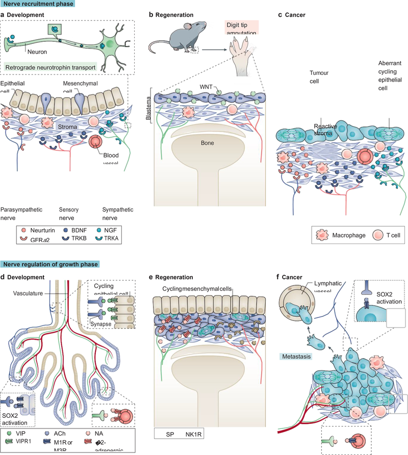
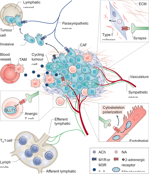
The contribution of nerves to the pathogenesis of malignancies has emerged as an important component of the tumour microenvironment. Recent studies have shown that peripheral nerves (sympathetic, parasympathetic and sensory) interact with tumour and stromal cells to promote the initiation and progression of a variety of solid and haematological malignancies. Furthermore, new evidence suggests that cancers may reactivate nerve-dependent developmental and regenerative processes to promote their growth and survival. Here we review emerging concepts and discuss the therapeutic implications of manipulating nerves and neural signalling for the prevention and treatment of cancer.


Conflict of interest statement

COMPETING INTERESTS

P.S.F. serves as consultant for Pfizer, has received research funding from Ironwood Pharmaceuticals and is shareholder of Cygnal Therapeutics. A.H.Z declares no competing interests.

PEER REVIEW INFORMATION

Nature Reviews Cancer thanks E. Repasky, S. Ben-Eliyahu and the other, anonymous, reviewer(s) for their contribution to the peer review of this work.

PUBLISHER’S NOTE

Springer Nature remains neutral with regard to jurisdictional claims in published maps and institutional affiliations.

Figures

Figure 1.
Figure 1.. Autonomic innervation of major primary sites of cancer formation in the mouse: anatomical targets for pre- and post-ganglionic surgical denervation.
This schematic depicts pre- and post-ganglionic innervation by the parasympathetic nervous system (PSNS), in purple, and the sympathetic nervous system (SNS), in green, of several well-studied organs where nerves have been implicated in disease progression. Anatomical landmarks are highlighted to indicate where pre- and post-ganglionic denervation of exocrine glands can occur. SNS outflow is depicted originating in the thoracic and lumbar regions of the spinal cord. The cell bodies of these preganglionic nerves (depicted by dotted lines) either project to the paravertebral sympathetic chain (the column of ganglia depicted just outside the spinal column) including the superior cervical and stellate ganglia, or project to distant ganglia including the celiac and hypogastric (pelvic) ganglia. Postganglionic nerves are depicted by solid lines. The salivary glands are a collection of three bilateral glands found in the mandibular region and consist of (1) the parotid gland, (2) the sublingual gland, and (3) the submaxillary gland. PSNS outflow is depicted originating in the brainstem and sacral region of the spinal cord. As depicted for the SNS, dotted lines represent preganglionic nerves, and solid lines represent postganglionic nerves.*, indicates sensory nuclei (nodose ganglion) whose projections are carried by the vagus nerve, along with parasympathetic fibers.
Figure 2.
Figure 2.. Reactivation of nerve-mediated developmental and regenerative pathways in cancer.
Nerve recruitment phase (a to c). During development, the epithelial, mesenchymal, and stromal compartments of organs secrete neurotrophins to recruit the three types of peripheral nerves (a). Neurotrophin binding to its cognate receptor on nerves leads to a signal that travels in a retrograde manner to the soma, affecting gene expression and axonal growth (inset). Similarly, in mammalian digit tip regeneration the stromal and mesenchymal compartments secrete neurotrophins (in this case, WNT) to recruit sympathetic and sensory nerves (b). In cancer, aberrantly mitotic malignant epithelial cells and the surrounding reactive stroma, release neurotrophins to recruit nerves necessary for their growth (c). Nerve-mediated regulation of the growth phase (d to f). During organ patterning, parasympathetic nerves initiate ductal epithelial cell (in beige) tubulogenesis (involving mitosis and migration) through vasoactive intestinal peptide (VIP) signalling (upper inset); parasympathetic nerves also regulate glandular or acinar epithelium (in purple) growth and patterning through acetylcholine (ACh) signalling by activating SRY-box 2 (SOX2) through a calcium-dependent pathway (lower left inset);,whereas sympathetic nerves pattern the vasculature (lower right inset) (d). During digit tip regeneration, sympathetic and sensory nerves provide mitotic and differentiation cues to the mesenchymal and overlying epithelial cells (e). Similarly, in cancer, sympathetic nerves pattern the neovasculature supplying the growing tumour, and parasympathetic nerves provide mitotic and migratory cues to tumour cells, which in turn lead to growth expansion and formation of micrometastases, respectively (f). BDNF, brain derived neurotrophic factor; GFRα2, glial cell line derived neurotrophic factor family receptor α2; NA, noradrenaline; NGF, nerve growth factor; NK1R, neurokinin 1 receptor; SP, substance P; TRK, tropomyosin receptor kinase.
Figure 3.
Figure 3.. Neural regulation of the tumour microenvironment.
Nerves interact with multiple stromal and malignant epithelial components to promote tumour growth and dissemination. The tumour microenvironment (TME) is largely immunosuppressive. Signalling from adrenergic nerves stimulates secretion of interleukin-8 (IL-8) from tumour cells, which then recruits tumour-associated macrophages (TAMs) that contribute to angiogenesis and further immunosuppression. Adrenergic nerves also signal to T cells through the β2-adrenergic receptor, suppressing metabolic activation by inhibiting expression of the glucose transporter GLUT1, and thus maintaining them in an anergic state (see inset). While immune-responsive T helper 1 (TH1) cells can be recruited to the tumour, they are often hindered from reaching it as increased adrenergic signalling to the lymphatic endothelium constricts the efferent lymphatic channels, thus trapping these cells in nearby lymph nodes.Angiogenesis, a key component of tumour development is intimately regulated by nerves. Signalling through endothelial-expressed β2-adrenergic receptor suppresses oxidative metabolism and promotes endothelial cell migration and vessel formation (see inset). In addition, cancer associated fibroblasts (CAFs) remodel the extracellular matrix through production of type I collagen in response to noradrenaline (NA) signalling (see inset). As mentioned in Figure 2, parasympathetic signalling through tumour cell-expressed cholinergic receptors, promotes tumour cell migration and formation of micrometastases. ACh, acetylcholine; M1R, muscarinic acetylcholine receptor 1.

References

    1. Hanahan D & Weinberg RA Hallmarks of cancer: the next generation. Cell 144, 646–674, doi:10.1016/j.cell.2011.02.013 (2011). - DOI - PubMed
    1. Zahalka AH et al. Adrenergic nerves activate an angio-metabolic switch in prostate cancer. Science 358, 321–326, doi:10.1126/science.aah5072 (2017).

      This paper shows that adrenergic nerves regulate the vasculature in the TME to promote tumour growth and cancer progression.

    1. Zhao CM et al. Denervation suppresses gastric tumorigenesis. Sci Transl Med 6, 250ra115, doi:10.1126/scitranslmed.3009569 (2014).

      This paper shows that surgical transection of the vagus nerve inhibits development of gastric cancer.

    1. Renz BW et al. beta2 Adrenergic-Neurotrophin Feedforward Loop Promotes Pancreatic Cancer. Cancer Cell, doi:10.1016/j.ccell.2017.11.007 (2017). - DOI - PMC - PubMed
    1. Magnon C et al. Autonomic nerve development contributes to prostate cancer progression. Science 341, 1236361, doi:10.1126/science.1236361 (2013).

      This paper showed a role for adrenergic and cholinergic nerves in prostate tumour growth and metastasis.

Publication types

Substances