. 2020 May;57(5):2194-2205.
doi: 10.1007/s12035-020-01867-9. Epub 2020 Jan 23.

Defining a Time Window for Neuroprotection and Glia Modulation by Caffeine After Neonatal Hypoxia-Ischaemia

Affiliations

Defining a Time Window for Neuroprotection and Glia Modulation by Caffeine After Neonatal Hypoxia-Ischaemia

Elena Di Martino et al. Mol Neurobiol. 2020 May.

Abstract

Hypoxic-ischemic (HI) brain injury remains an important cause of brain damage in neonates with potential life-long consequences. Caffeine, which is a competitive inhibitor of adenosine receptors, is commonly used as treatment for preterm apnoea in clinical settings. In the current study, we investigated the effects of caffeine given at 0 h, 6 h, 12 h or 24 h after HI in P10 mouse pups. Open field and rotarod behavioural tests were performed 2 weeks after injury, and brain morphology was then evaluated. Gene expression and immunohistological analyses were assessed in mice 1- and 5-day post-HI. A single dose of caffeine directly after HI resulted in a reduction of the lesion in the grey and white matter, judged by immunostaining of MAP2 and MBP, respectively, compared to PBS-treated controls. In addition, the number of amoeboid microglia and apoptotic cells, the area covered by astrogliosis, and the expression of pro-inflammatory cytokines were significantly decreased. Behavioural assessment after 2 weeks showed increased open-field activity after HI, and this was normalised if caffeine was administered immediately after the injury. Later administrations of caffeine did not change the outcomes when compared to the vehicle group. In conclusion, caffeine only yielded neuroprotection and immunomodulation in a neonatal model of brain hypoxia ischaemia if administered immediately after injury.

Keywords: Caffeine; Hypoxia-ischemia; Immunomodulation; Neuroprotection; Time-window.


Figures

Fig. 1
Fig. 1
Long-term evaluation of caffeine treatment (P27). Mice were subjected to HI and received PBS (n = 8) or caffeine (n = 10) acutely (0 h) or at either 6 h (n = 6), 12 h (n = 6) and 24 h (n = 7) after injury. Neuropathological score in sections stained with cresyl violet (a), tissue loss calculated by MAP2-stained tissue (b), third day activity in the open field test (c) and rotarod (d) behavioural performances 2 weeks after HI. The dashed-lines in figures (c) and (d) represent the performance of sham animals as an internal control. Data are presented as mean ± SEM with *p < 0.05 (two-way ANOVA with Dunnet’s multiple comparison test for repeated measurements for open-field test and one-way ANOVA with Bonferroni’s multiple comparison test for the other graphs)
Fig. 2
Fig. 2
Short-term evaluation of caffeine treatment (P15). Mice were subjected to HI and received caffeine (n = 10) or PBS (n = 8) acutely after injury (0 h). Neuropathological score in sections stained with cresyl violet (a), tissue loss calculated by MAP2-stained tissue at the level of the striatum and hippocampus (b). Example of overlapping areas between the tissue loss (MAP2) and astrogliosis (GFAP) after HI injury (c). Glial scar percentage calculated by GFAP staining in different brain regions (d) and an example of reactive astrocytes contouring at × 10 with scale bar 100 μm (e). Data are presented as mean ± SEM with *p < 0.05 and **p < 0.01 (unpaired t test)
Fig. 3
Fig. 3
Analysis of apoptotic cells using TUNEL staining. Caffeine reduced the number of apoptotic cells in the cortex (a) and striatum (b) of mice after HI (n = 10) if compared to the PBS group (n = 8). Example of TUNEL+ cells at × 40 with scale bar 50 μm in the small squares and representative tiled-images of the cortex (Cx) and striatum (Str) at × 10 with scale bar 300 μm in the big squares (c). Data are presented as mean ± SEM with *p < 0.05 (unpaired t test)
Fig. 4
Fig. 4
Caffeine modulates microglial cells’ density and morphology. The density of Iba1+ amoeboid microglia was reduced in the cortex (a) and striatum (b) of mice receiving caffeine (n = 10) compared to the PBS group (n = 8). The area of the soma of phagocytic Iba1+ cells was reduced after caffeine treatment in the cortex (c). Sham animals receiving caffeine (n = 4) did not show any alteration of microglia numbers or shape when compared to the Sham + PBS group (n = 6). Example of Iba1 staining at × 40 with scale bar 30 μm (d) and representative images of the cortices at × 20 with scale bar 50 μm (e). Data are presented as mean ± SEM with *p < 0.05 and **p < 0.01 (two-way ANOVA with Sidak’s multiple comparison test)
Fig. 5
Fig. 5
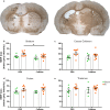
Caffeine prevents white matter injury in the striatum. Myelin basic protein (MBP) density was analysed in four different brain areas, namely the cortex (Cx), striatum (Str), corpus callosum (CC) and thalamus (Th) as indicated from the squares (a). HI injury led to a significant loss of myelination in almost all the analysed brain areas. Injured mice receiving caffeine (n = 10) showed a less-extensive MBP loss in the striatum (b) but not observable differences were found in the corpus callosum (c), cortex (d) or thalamus (e) if compared to PBS group (n = 8). Sham animals receiving caffeine (n = 4) did not show any anomaly compared to the sham mice receiving PBS (n = 6). Data are presented as mean ± SEM with *p < 0.05 (two-way ANOVA with Sidak’s multiple comparison test)
Fig. 6
Fig. 6
Caffeine prevents upregulation of the expression of pro-inflammatory genes 24 h after HI. RT-qPCR analysis of Il1b, Il6, Il12 and Ifng genes in brain homogenates derived from ipsilateral cortices of HI mice receiving either caffeine (n = 13) or PBS (n = 12) normalised to control animals. The expression of each gene was calculated by normalisation to the average of housekeeping genes B-actin and Rpl13a. Data are presented as mean ± SEM with *p < 0.05 (unpaired t test)

References

    1. Victora CG, Requejo JH, Barros AJ, Berman P, Bhutta Z, Boerma T, Chopra M, De Francisco A, Daelmans B, Hazel E. Countdown to 2015: a decade of tracking progress for maternal, newborn, and child survival. Lancet. 2016;387(10032):2049–2059. doi: 10.1016/S0140-6736(15)00519-X. - DOI - PMC - PubMed
    1. Volpe JJ. Perinatal brain injury: from pathogenesis to neuroprotection. Ment Retard Dev Disabil Res Rev. 2001;7(1):56–64. doi: 10.1002/1098-2779(200102)7:1<56::Aid-mrdd1008>3.0.Co;2-a. - DOI - PubMed
    1. Wassink G, Davidson JO, Lear CA, Juul SE, Northington F, Bennet L, Gunn AJ (2018) A working model for hypothermic neuroprotection. J Physiol. 10.1113/jp274928 - PMC - PubMed
    1. Robertson NJ, Nakakeeto M, Hagmann C, Cowan FM, Acolet D, Iwata O, Allen E, Elbourne D, Costello A, Jacobs I. Therapeutic hypothermia for birth asphyxia in low-resource settings: a pilot randomised controlled trial. Lancet (London, England) 2008;372(9641):801–803. doi: 10.1016/s0140-6736(08)61329-x. - DOI - PubMed
    1. Fredholm BB, AP IJ, Jacobson KA, Linden J, Muller CE. International Union of Basic and Clinical Pharmacology. LXXXI Nomenclature and classification of adenosine receptors--an update. Pharmacol Rev. 2011;63(1):1–34. doi: 10.1124/pr.110.003285. - DOI - PMC - PubMed

MeSH terms