Review
. 2020 Jun 24;120(12):4921-4968.
doi: 10.1021/acs.chemrev.9b00489. Epub 2020 Jan 24.

Biosynthesis of Nitrogenase Cofactors

Affiliations
Review

Biosynthesis of Nitrogenase Cofactors

Stefan Burén et al. Chem Rev. .

Abstract

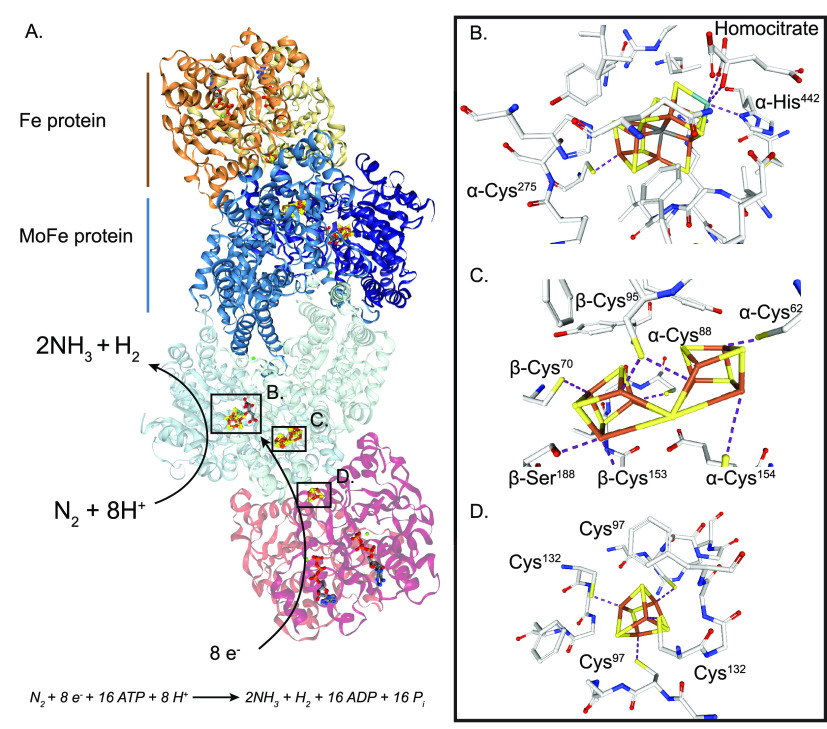
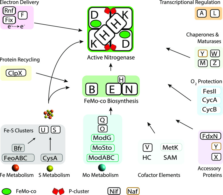
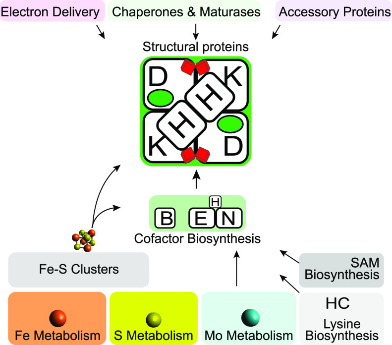
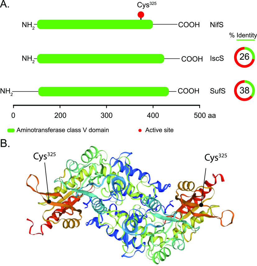
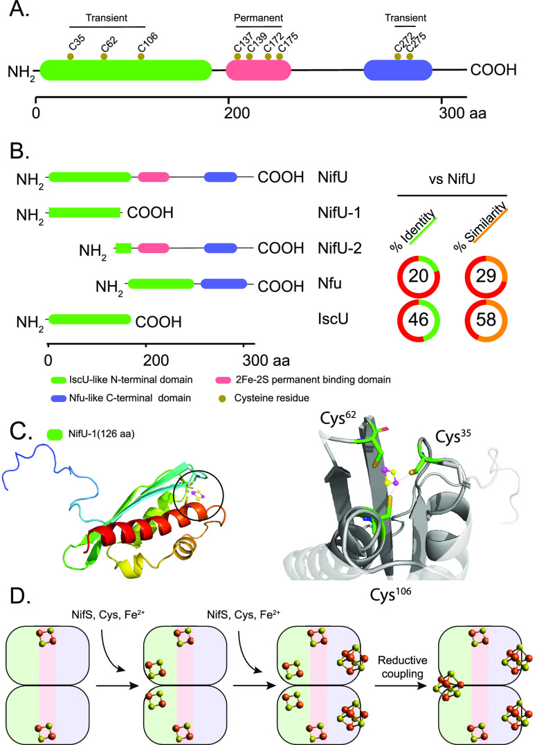
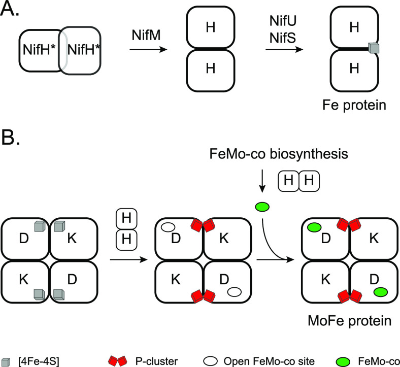
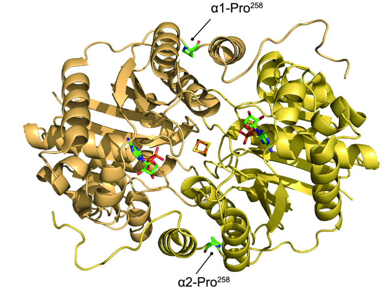
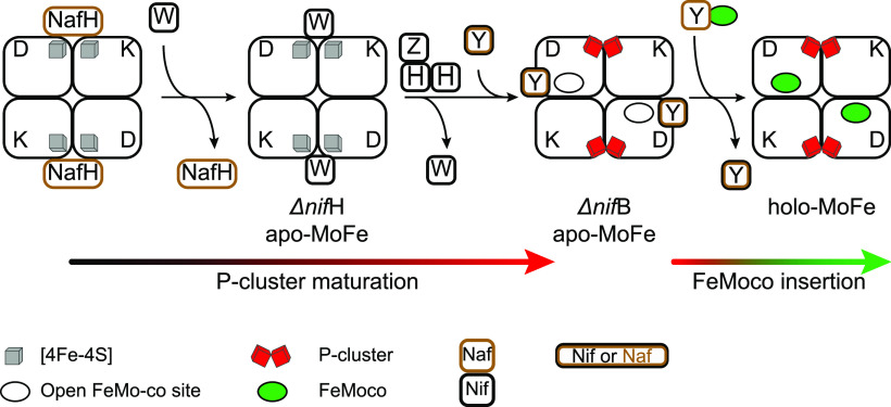
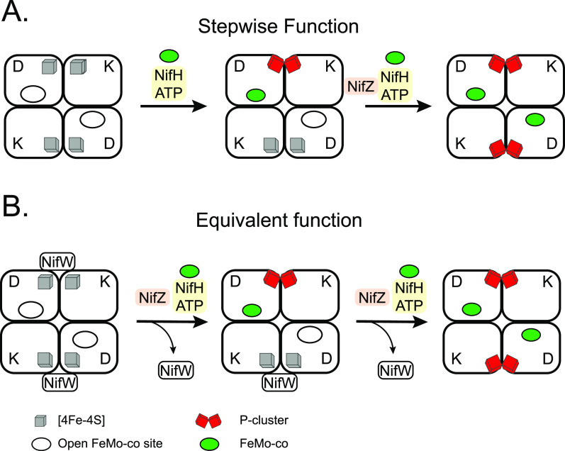
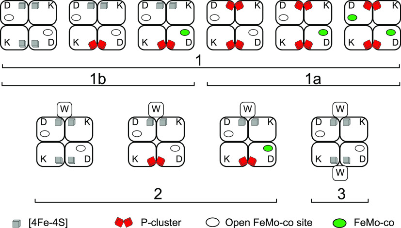
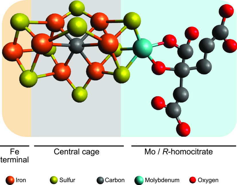
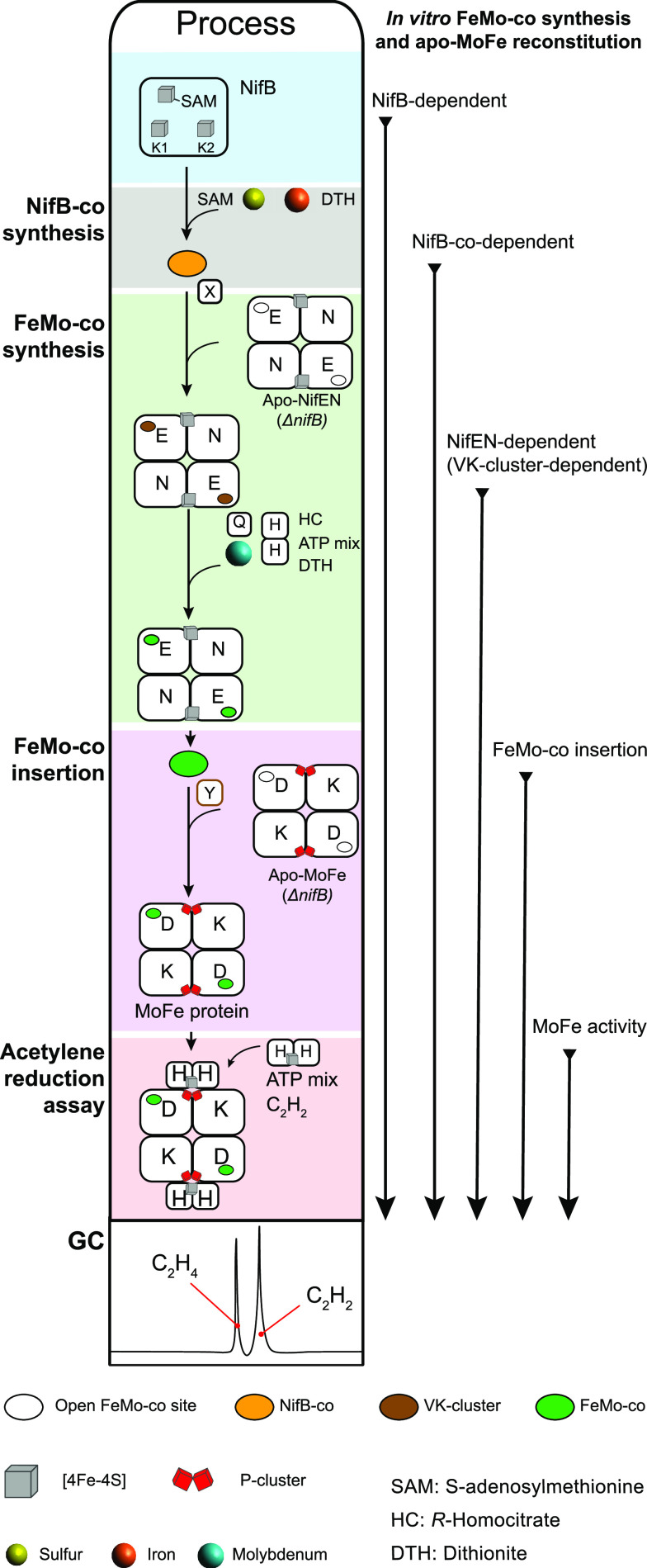
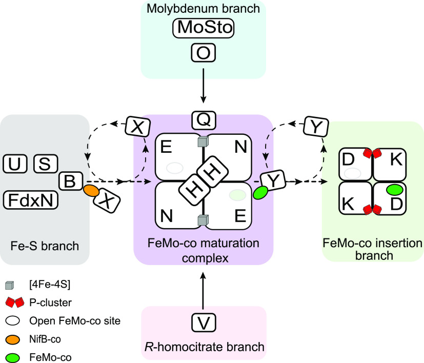
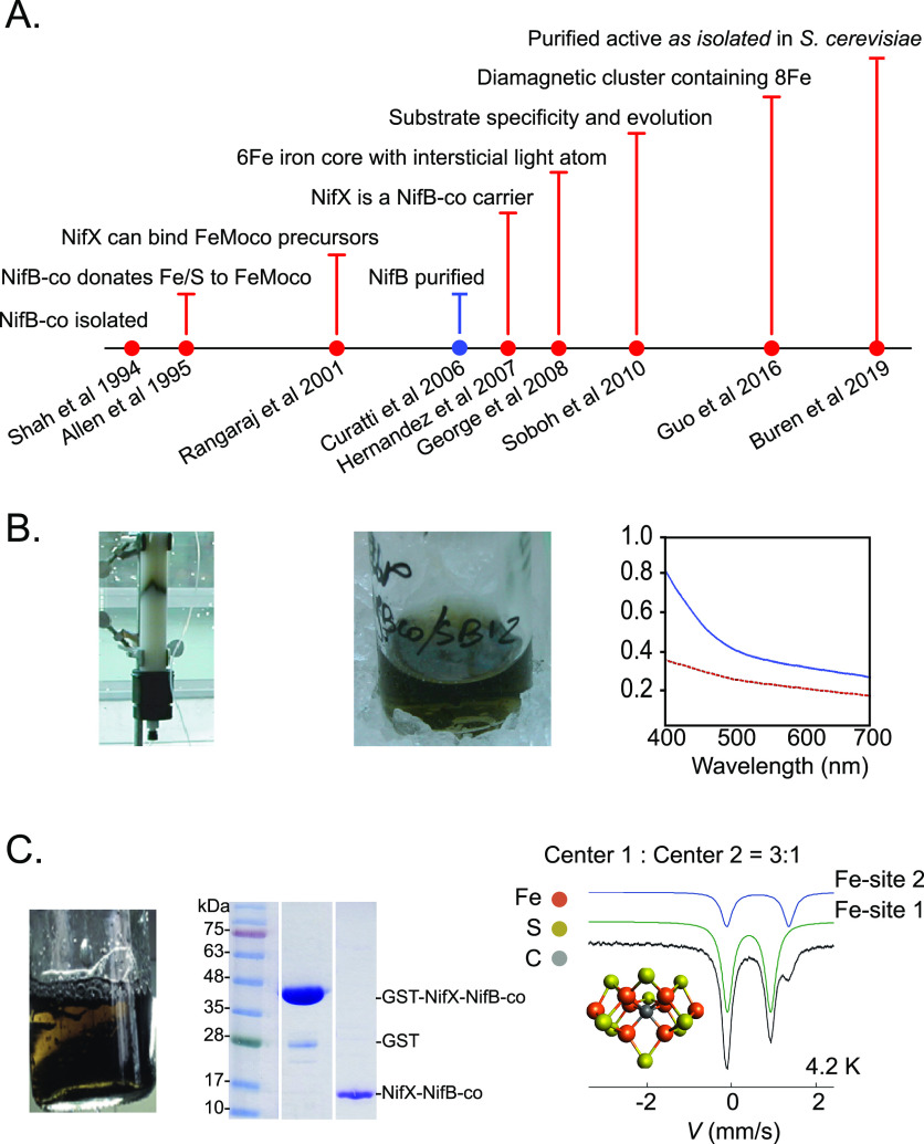
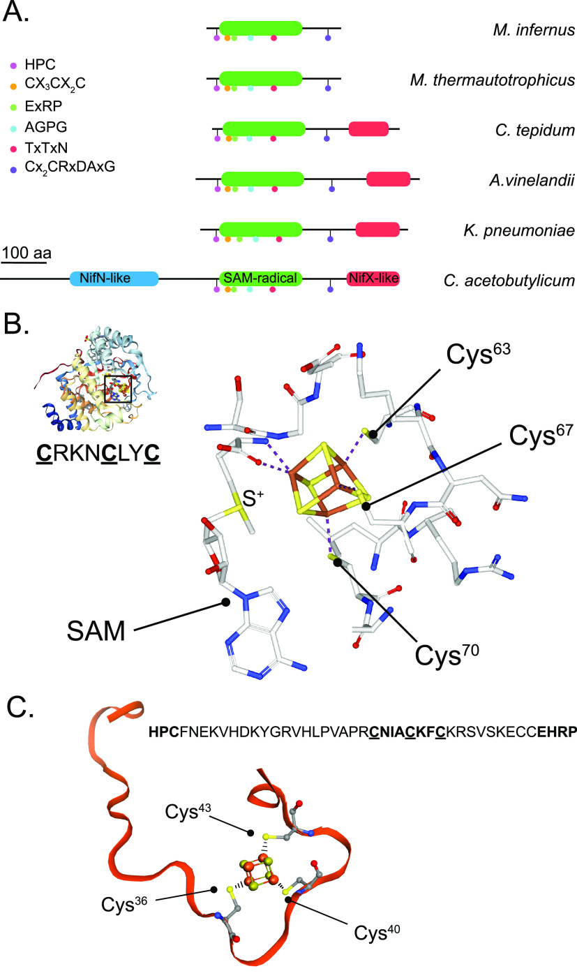
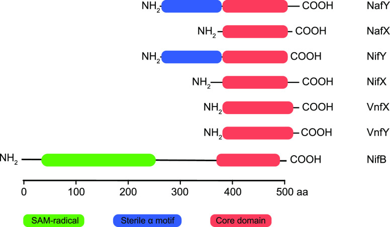
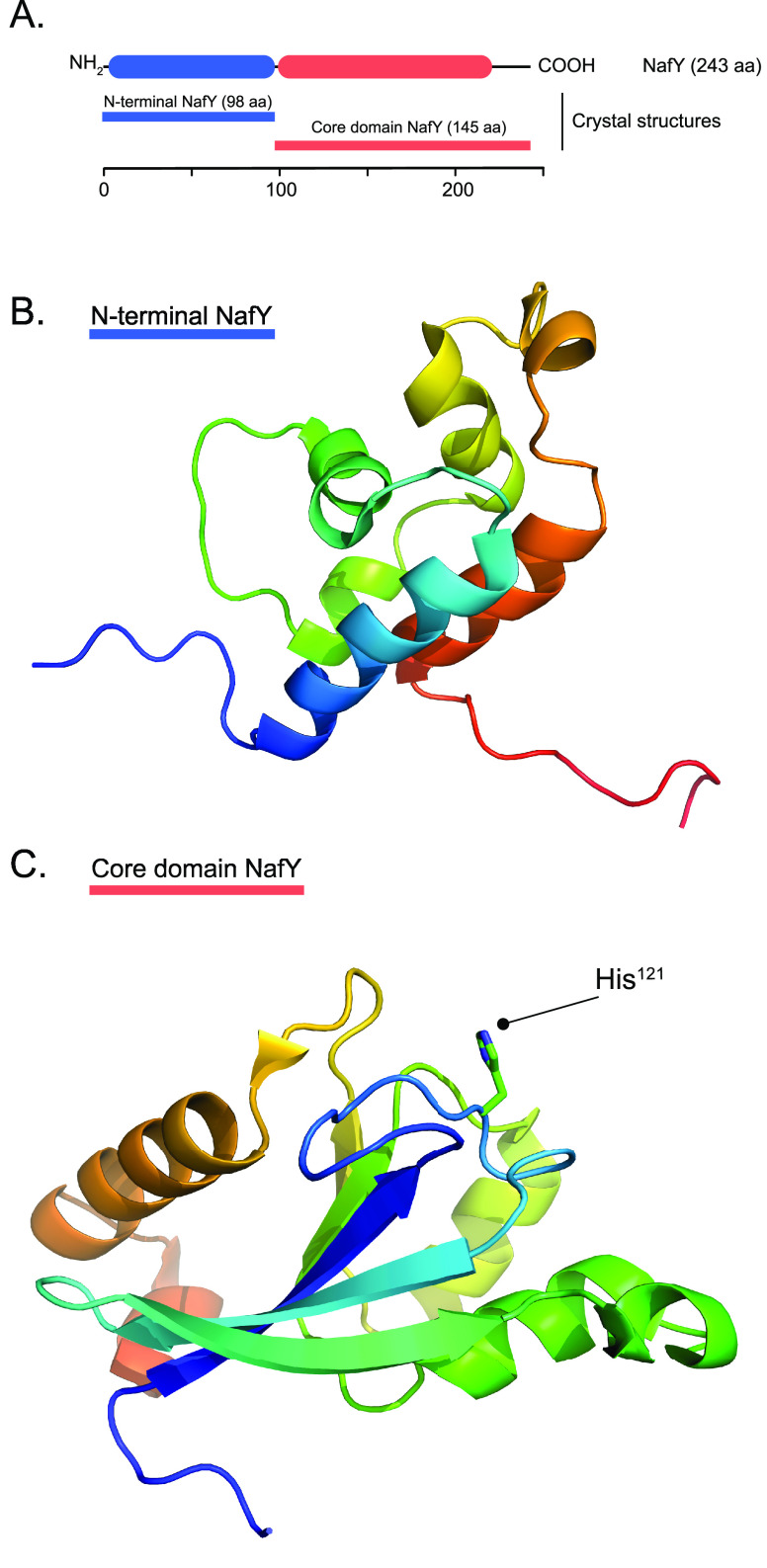
Nitrogenase harbors three distinct metal prosthetic groups that are required for its activity. The simplest one is a [4Fe-4S] cluster located at the Fe protein nitrogenase component. The MoFe protein component carries an [8Fe-7S] group called P-cluster and a [7Fe-9S-C-Mo-R-homocitrate] group called FeMo-co. Formation of nitrogenase metalloclusters requires the participation of the structural nitrogenase components and many accessory proteins, and occurs both in situ, for the P-cluster, and in external assembly sites for FeMo-co. The biosynthesis of FeMo-co is performed stepwise and involves molecular scaffolds, metallochaperones, radical chemistry, and novel and unique biosynthetic intermediates. This review provides a critical overview of discoveries on nitrogenase cofactor structure, function, and activity over the last four decades.


Conflict of interest statement

The authors declare no competing financial interest.

Figures

Figure 1
Figure 1
Structure of Mo nitrogenase. (A) A. vinelandii MoFe protein and Fe protein complex (PDB: 1G21). Top half shows polypeptide secondary structure. Partial transparency has been applied to the polypeptide chains of the bottom half to visualize metal clusters and nucleotides. Ligands and surrounding environment for (B) FeMo-co, (C) P-cluster and (D) [4Fe-4S] cluster are shown. Mo nitrogenase reaction is shown at the bottom. Images created with NGL viewer and RCSB PDB.
Figure 2
Figure 2
Organization and proposed functions of Mo nitrogenase genes in A. vinelandii. Figure shows the chromosomal location and genetic organization of the major and minor nif gene clusters. Numbers to the left and right of each gene cluster indicate chromosomal location. Transcriptional units are depicted by arrows. Proposed roles in Mo nitrogenase are color coded in the legend. Metal clusters embedded in Mo nitrogenase and a surface structure of the complex are shown.
Figure 3
Figure 3
Processes for Mo-nitrogenase biogenesis in the model bacterium A. vinelandii. Proteins involved in each process are shown.
Figure 4
Figure 4
Simplification of genetic requirements for Mo-nitrogenase biogenesis achieved by its integration with housekeeping processes. The essential six gene core is highlighted in green in the middle. Housekeeping processes provide the functions of nitrogenase ancillary proteins.
Figure 5
Figure 5
A. vinelandii NifS cysteine desulfurase. (A) Domain composition and % identity of A. vinelandii NifS (Uniprot C1DH19) compared to IscS (Uniprot C1DH19) and SufS (Uniprot C1DH19). (B) Structure of the NifS homologue protein CsdB showing the surface position of the active-site Cys residue. Image created with NGL viewer and RCSB PDB.
Figure 6
Figure 6
Modularity of NifU structure and function. (A) A. vinelandii NifU domain architecture and conserved Cys residues (Uniprot C1DH18). Three distinct NifU domains are shown in green, pink, and blue, and their roles in coordinating permanent or transient [Fe–S] clusters are indicated. (B) Alignment of full-length NifU with the NifU-1 and NifU-2 truncated variants and with the homologous proteins IscU (Uniprot C1DE67) and human NfU (Uniprot C1DLW0). Conserved domains are color coded. % Identity and similarity between NifU and NfU or IscU are shown to the right. (C). Structural model of the N-terminal domain of NifU generated with Swiss-Model. The protein pocket with ligands to a transient [2Fe-2S] cluster is magnified to the right of the structure. Graphics generated with the PyMOL Molecular Graphics System, Version 2.3.2 Schrödinger, LLC. (D) Proposed model for NifS-mediated assembly of [4Fe-4S] clusters in the N-terminal and C-terminal domains of the NifU.
Figure 7
Figure 7
Fe protein maturation and the roles of the Fe protein in MoFe protein maturation. (A) Simplified model for the roles of NifM, NifU and NifS in Fe protein maturation. (B) Requirement of Fe protein for MoFe protein maturation. MoFe protein variants from left to right correspond to the ΔnifH apo-MoFe protein, the ΔnifB apo-MoFe protein, and the holo-MoFe protein. Fe protein is shown as NifH homodimer lacking [4Fe-4S] cluster to indicate that apo-Fe protein is competent in P-cluster formation and in FeMo-co biosynthesis.
Figure 8
Figure 8
Location of Pro258 in a top-view structure of the A. vinelandii Fe protein (PDB 1FP6). The two monomers, the central [4Fe-4S] cluster, and the two nucleotides are easily identified. Graphics generated with the PyMOL Molecular Graphics System, Version 2.3.2 Schrödinger, LLC.
Figure 9
Figure 9
Sequential and differential interaction of NafH, NifW and NifZ maturation factors with apo-MoFe protein. From left to right, three steps of MoFe protein maturation related to P-cluster formation and one step for the insertion of FeMo-co are shown. NifZ aids the Fe protein in P-cluster formation. NafH and NifW interaction with the MoFe protein precedes the NifH/NifZ reaction. Formation of the P-clusters releases NifW, changes apo-MoFe protein conformation to make the FeMo-co sites accessible, and promotes the binding of the NifY or NafY factors. FeMo-co insertion releases NifY/NafY and generates holo-MoFe protein.
Figure 10
Figure 10
Models for the participation of NifZ in P-cluster maturation. (A) In the stepwise model, the first P-cluster of each tetramer is matured by the Fe protein alone, while both NifZ and the Fe protein are essential for the maturation of the second P-cluster. (B) In the equivalent function model, NifZ participates in the formation of both P-clusters, but it is not essential for either process. In both models, ATP is required for P-cluster formation. The equivalent model incorporates information about the release of NifW upon P-cluster maturation. Figure adapted from ref (116). Copyright 2019 ASBMB under CC BY 4.0 http://creativecommons.org/licenses/by/4.0/.
Figure 11
Figure 11
Theoretical apo-MoFe variants and holo-MoFe protein that could accumulate in an A. vinelandii ΔnifZ strain. Chromatography fractions and subfractions in which these proteins were identified are number as 1, 1a, 1b, 2, and 3 (see text). Legend indicates the presence of [4Fe-4S] P-cluster precursors, FeMo-co, and the NifW maturation factor. Figure adapted from ref (116). Copyright 2019 ASBMB under CC BY 4.0 http://creativecommons.org/licenses/by/4.0/.
Figure 12
Figure 12
Structure of FeMo-co. The structure shows the trigonal prism at the center of the cofactor, the capping Fe and Mo atoms, and the location of R-homocitrate.
Figure 13
Figure 13
Assays for in vitro FeMo-co synthesis and apo-MoFe protein reconstitution. This schematic diagram shows the most relevant in vitro FeMo-co synthesis assays described in the literature and labeled to the right as NifB-dependent, NifB-co-dependent, NifEN-dependent, FeMo-co insertion, and MoFe protein activity, relating to the starting component (protein or cluster precursor) being tested. The assays can be performed by mixing cell extracts containing the indicated components or by using highly purified proteins, precursors and chemicals. The most complex assay would be the NifB-dependent (requires competent NifB containing its [Fe–S] cluster complement), which includes NifB, SAM, ΔnifB apo-NifEN, Na2MoO4, homocitrate, Fe protein, DTH, ΔnifB apo-MoFe protein, and ATP mix. NifX and NafY are not essential but stimulate FeMo-co synthesis and insertion. Na2MoO4 can be replaced by NifQ.
Figure 14
Figure 14
Simplified model for FeMo-co biosynthesis centered in the hub formed by NifEN and the Fe protein designated in the figure as FeMo-co maturation complex. The pathways providing the FeMo-co Fe–S cage, molybdate, and homocitrate are shown and include the proteins involved. Roles of NifX and NafY in delivering NifB-co and FeMo-co to NifEN and the MoFe protein, respectively, are indicated.
Figure 15
Figure 15
NifB-co is a diamagnetic [8Fe-9S–C] cluster. (A) Timeline showing the milestones from the discovery of NifB-co to its final characterization together with the first author and year of the reports. (B) NifB-co as isolated from membranes of K. pneumoniae. NifB-co solutions are greenish brown with nondescript visible spectra in the 400–700 nm region. (C) Isolation of an in vivo formed NifX-NifB-co complex and its Mössbauer analysis revealing two spectroscopically different Fe sites at 3:1 ratio. UV–visible spectrum reprinted with permission from ref (19). Copyright 1994 ASBMB. Mössbauer spectra adapted with permission from ref (20). Copyright 2016 John Wiley and Sons.
Figure 16
Figure 16
(A) NifB domain architectures and conserved amino acid motifs. The simplest NifB proteins consist of a stand-alone SAM radical motif. More complex NifB proteins include a C-terminal NifX-like, or N-terminal NifN-like domain and C-terminal NifX-like domain. (B) Structure of the NifB homologue MoaA showing the [4Fe-4S]-SAM active site and its ligating Cys residues. Images created with NGL viewer and RCSB PDB. No NifB crystal structure is yet available. (C) Structural model, generated with SWISS-MODEL, of a 43 amino acid fragment of the A. vinelandii NifB sequence including the AdoMet motif Cx3Cx2C.
Figure 17
Figure 17
NifB phylogeny and distribution of NifB domain architectures among putative diazotrophs. The tree shows overall distribution and relative frequency of NifB architectures. Figure reprinted from ref (64). Copyright 2017 Arragain, Jiménez-Vicente, Scandurra, Burén, Rubio, and Echavarri-Erasun under CC BY 4.0 http://creativecommons.org/licenses/by/4.0/.
Figure 18
Figure 18
Spectroscopic signals from NifB [Fe–S] clusters. (A) SDS gel of purified M. infernus NifB used for spectroscopic analyses. (B) UV–vis spectra of as-isolated and Fe–S cluster reconstituted NifB variants. All spectra were recorded in DTH-reduced samples. (C) Fe content and Fe–S cluster assignment for the NifB variants. (D) Deconvolution of the EPR signal arising from the three distinct [4Fe-4S] clusters of NifB. Recorded data are shown in black and the simulations in blue. The signal from the RS ([4Fe-4S]-SAM cluster) component is shown in red, that of the K1 component in blue, and that of the K2 component in pink. Figure adapted from ref (58). Copyright 2016 ACS.
Figure 19
Figure 19
Model of the mechanism of NifB-co formation by NifB. (A) Proposed steps based on biochemical evidence: the K1 cluster presents mixed Cys-His coordination; SAM-dependent methyl group transfer to the K2 cluster; reductive cleavage of a second SAM molecule and formation of a 5′-dA radical at the active site; methyl group H atom abstraction by the 5′-dA radical; oxidation/deprotonation events to remove to H atoms; core rearrangement and insertion of a S atom at the belt of the precursor. (B) Hypothesized role of FdxN in reduction of the K2 cluster to prepare it for methyl group acceptance.
Figure 20
Figure 20
Mo uptake, storage, and processing for nitrogenase in A. vinelandii. (A) Scheme summarizing current understanding of molybdenum trafficking in A. vinelandii. Molybdate in the medium is chelated by siderophores protochelin and azotochelin. Internalization to the periplasm space is believed to occur via outer membrane porins. Active transport of molybdate (and tungstate) to the cytoplasm is mediated by ABC transporters composed of ModABC polypeptides. Most molybdate in A. vinelandii is stored at MoSto complex in a process energized by ATP hydrolysis. However, in Mo-starved A. vinelandii cells, MoSto can store tungstate forming WSto. Mo in MoSto is available to the molybdoenzymes nitrogenase and nitrate reductase. Pathway branching appears to occur before involvement of ModG, a trimer that binds eight molybdates at the interface of its subunits. NifO is also involved in directing Mo toward FeMo-co against the Mo-co branch. NifQ carries a [Mo-3Fe-4S] cluster shown to deliver Mo to the NifEN scaffold protein where it will be incorporated into an Fe–S cluster precursor to generate FeMo-co. The structures of a ModA2B2C2 transporter (PDB: 2ONK), the α3β3 MoSto (PDB: 6GU5) and WSto (PDB: 2OGX), an α3 ModG (PDB 1H9M), α2β2 NifEN (PDB: 3PDI), and the Fe protein (PDB: 1NIP) are shown. Structure images created with NGL viewer and RCSB PDB. (B) Molybdenum and tungsten electronic shells and their molybdate and tungstate forms. (C) Common A. vinelandii metalophores. (D) Inside details of the MoSto protein crystal structure shown in panel A including POMs (molybdenum-based polyoxometalates) and ATP sites.
Figure 21
Figure 21
Metal cluster interconversions in NifQ. NifQ preparations carry substoichiometric amounts of [Mo-3Fe-4S-2O] cluster (0.3 mol cluster per mol NifQ) and an independent MoO2S2 species. A [3Fe-4S] cluster species makes up the full cluster complement (0.7 mol cluster per mol NifQ). Redox-gated incorporation of Mo into the [3Fe-4S] cluster has been shown. Molybdenum bound to NifQ is delivered to a NifEN/Fe protein complex for FeMo-co biosynthesis.
Figure 22
Figure 22
Coordination around homocitrate in the A. vinelandii MoFe protein (PDB: 1M1N). (A) Depiction of the FeMo-co pocket showing solvent accessible tunnels to the cofactor. (B) Network of hydrogen bonds among homocitrate, nearby amino acid residues, and the pool of water molecules inside the pocket. (C) Interactions of the Mo atom with His442 and with the C2 hydroxyl and carboxyl groups of homocitrate. Images created with NGL viewer and RCSB PDB.
Figure 23
Figure 23
Comparison of NifEN and MoFe protein structures. (A) Cartoon depiction of NifEN atomic structure (PDB: 3PDI). Partial transparency has been applied to the polypeptide chains to visualize metal clusters. (B) Cartoon depiction of MoFe protein atomic structure (PDB: 3K1A). (C) [4Fe-4S] cluster structure and coordination by Cys residues in NifEN. (D) VK-cluster structure and coordination to α-Cys of NifEN. Note superficial position of the VK-cluster in the protein. Each αβ half of NifEN contains a [4Fe-4S] cluster and a VK-cluster. Graphics generated with the PyMOL Molecular Graphics System, Version 2.3.2 Schrödinger, LLC.
Figure 24
Figure 24
Position in the A. vinelandii Fe protein of amino acid residues (from Table 3) mutated to investigate their involvement in Fe protein function. (A) Semi transparent surface and loop representation. (B) Ribbon diagram in the same orientation. (C) Main chain loop representation. Graphics generated with the PyMOL Molecular Graphics System, Version 2.3.2 Schrödinger, LLC. ADP molecules are represented as filled spheres at the bottom part of the protein. The [4Fe-4S] cluster is depicted with stick and balls in the upper part of the protein. Each mutated amino acid residue is assigned a different color.
Figure 25
Figure 25
Two models for NifEN metal cluster composition and Mo incorporation into an Fe–S cluster FeMo-co precursor to mature the cofactor within NifEN. Each model shows the protein and metal cluster nomenclature used. EPR signals used as spectroscopic evidence of protein-bound FeMo-co, FeMo-co precursors, and other MoFeS clusters are shown. (A) Model in which the product of NifB, called L-cluster, is delivered by NifB to apo-NifEN via direct interaction to generate the NifENL form. In subsequent step, the Fe protein delivers a Mo-homocitrate complex that replaces one terminal Fe atom in the L-cluster to generate the M-cluster found in a NifENM species. The M-cluster is transferred from NifENM to apo-MoFe protein by direct interaction to generate active MoFe protein. (B) Model in which the product of NifB, called NifB-co, is transferred to apo-NifEN via NifX. Within NifEN, NifB-co is rapidly transformed into the VK-cluster by NifEN. VK-cluster can be extracted from NifEN by NifX. Both NifX/NifB-co and NifX/VK-cluster complexes have been spectroscopically analyzed. In addition to the VK-cluster, NifEN contains a [Mo-3Fe-4S] cluster proposed to be donated by NifQ, which carries a similar cluster. Homocitrate is donated by NifV. Once all FeMo-co precursors are bound to NifEN, the docked Fe protein exerts a conformational change in NifEN that promotes FeMo-co formation. Mature FeMo-co is transferred to apo-MoFe protein via NafY. NifQ inset adapted from ref (73). Copyright 2008 PNAS.
Figure 26
Figure 26
Core domain conservation among members of NafY family of proteins. NafY and homologues shown are from A. vinelandii. UniProt accession numbers are NafY (C1DMA1), NifY (C1DH00), NifX (C1DH05), VnfX (C1DI38), VnfY (C1DI21), and NifB (C1DMB1). Core domain is shown in orange, SAM-radical domain in green, and the sterile α motif domain in blue.
Figure 27
Figure 27
Atomic structures of NafY N-terminal and core domains. (A) N-terminal and C-terminal (core) domains of NafY. (B) NafY N-terminal domain structure (98 residues) solved by NMR (PDB 2KIC). (C) NafY core domain structure (145 residues) solved by X-ray crystallography (PDB 1P90). The His residue essential to FeMo-co binding is labeled. Graphics generated with the PyMOL Molecular Graphics System, Version 2.3.2 Schrödinger, LLC.

References

    1. Hoffman B. M.; Lukoyanov D.; Yang Z.-Y. Y.; Dean D. R.; Seefeldt L. C. Mechanism of Nitrogen Fixation by Nitrogenase: The Next Stage. Chem. Rev. 2014, 114, 4041–4062. 10.1021/cr400641x. - DOI - PMC - PubMed
    1. Bulen W. A.; LeComte J. R. The Nitrogenase System from Azotobacter: Two-Enzyme Requirement for N2 Reduction, ATP-Dependent H2 Evolution, and ATP Hydrolysis. Proc. Natl. Acad. Sci. U. S. A. 1966, 56, 979–986. 10.1073/pnas.56.3.979. - DOI - PMC - PubMed
    1. Hageman R. V.; Burris R. H. Nitrogenase and Nitrogenase Reductase Associate and Dissociate with Each Catalytic Cycle. Proc. Natl. Acad. Sci. U. S. A. 1978, 75, 2699–2702. 10.1073/pnas.75.6.2699. - DOI - PMC - PubMed
    1. Duval S.; Danyal K.; Shaw S.; Lytle A. K.; Dean D. R.; Hoffman B. M.; Antony E.; Seefeldt L. C. Electron Transfer Precedes ATP Hydrolysis during Nitrogenase Catalysis. Proc. Natl. Acad. Sci. U. S. A. 2013, 110, 16414–16419. 10.1073/pnas.1311218110. - DOI - PMC - PubMed
    1. Georgiadis M. M.; Komiya H.; Chakrabarti P.; Woo D.; Kornuc J. J.; Rees D. C. Crystallographic Structure of the Nitrogenase Iron Protein from Azotobacter Vinelandii. Science 1992, 257, 1653–1659. 10.1126/science.1529353. - DOI - PubMed

Publication types

MeSH terms