. 2019 Dec:71:141-245.
doi: 10.12905/0380.sydowia71-2019-0141. Epub 2019 Dec 23.

Fungal Systematics and Evolution: FUSE 5

Jie Song  1 Jun-Feng Liang  1 Mehdi Mehrabi-Koushki  2   3 Irmgard Krisai-Greilhuber  4 Barkat Ali  5   6 Vinod Kumar Bhatt  7 Agustín Cerna-Mendoza  8 Bin Chen  1 Zai-Xiong Chen  9 Hong-Long Chu  10 Mike Anderson Corazon-Guivin  8 Gladstone Alves da Silva  11 André De Kesel  12 Bálint Dima  13 Francesco Dovana  14 Reza Farokhinejad  2 Guliano Ferisin  15 Juan Carlos Guerrero-Abad  8   16 Ting Guo  17 Li-Hong Han  10 Sobia Ilyas  18 Alfredo Justo  19 Abdul Nasir Khalid  20 Sadigheh Khodadadi-Pourarpanahi  2 Tai-Hui Li  21 Chao Liu  10 Marilinda Lorenzini  22 Jun-Kun Lu  1 Abdul Samad Mumtaz  5 Fritz Oehl  23 Xue-Yu Pan  1 Viktor Papp  24 Wu Qian  25 Abdul Razaq  26 Kamal C Semwal  27 Li-Zhou Tang  10 Xue-Lian Tian  10 Adela Vallejos-Tapullima  8 Nicolaas A van der Merwe  6 Sheng-Kun Wang  1 Chao-Qun Wang  21 Rui-Heng Yang  17 Fei Yu  1 Giacomo Zapparoli  22 Ming Zhang  21 Vladimir Antonín  28 André Aptroot  29 Ali Aslan  30 Arghya Banerjee  31 Subrata Chatterjee  32 Alden C Dirks  33 Leila Ebrahimi  34 Khalil-Berdi Fotouhifar  35 Youbert Ghosta  36 Lyudmila B Kalinina  37 Dilara Karahan  38 Jingyu Liu  39 Mrinal Kumar Maiti  40 Abhirup Mookherjee  40 Partha Sarathi Nath  31 Birendranath Panja  31 Jayanta Saha  31 Hana Ševčíková  28 Hermann Voglmayr  4   41 Kenan Yazıcı  38 Danny Haelewaters  39   42   43   44
Affiliations

Fungal Systematics and Evolution: FUSE 5

Jie Song et al. Sydowia. 2019 Dec.

Abstract

Thirteen new species are formally described: Cortinarius brunneocarpus from Pakistan, C. lilacinoarmillatus from India, Curvularia khuzestanica on Atriplex lentiformis from Iran, Gloeocantharellus neoechinosporus from China, Laboulbenia bernaliana on species of Apenes, Apristus, and Philophuga (Coleoptera, Carabidae) from Nicaragua and Panama, L. oioveliicola on Oiovelia machadoi (Hemiptera, Veliidae) from Brazil, L. termiticola on Macrotermes subhyalinus (Blattodea, Termitidae) from the DR Congo, Pluteus cutefractus from Slovenia, Rhizoglomus variabile from Peru, Russula phloginea from China, Stagonosporopsis flacciduvarum on Vitis vinifera from Italy, Strobilomyces huangshanensis from China, Uromyces klotzschianus on Rumex dentatus subsp. klotzschianus from Pakistan. The following new records are reported: Alternaria calendulae on Calendula officinalis from India; A. tenuissima on apple and quince fruits from Iran; Candelariella oleaginescens from Turkey; Didymella americana and D. calidophila on Vitis vinifera from Italy; Lasiodiplodia theobromae causing tip blight of Dianella tasmanica 'variegata' from India; Marasmiellus subpruinosus from Madeira, Portugal, new for Macaronesia and Africa; Mycena albidolilacea, M. tenuispinosa, and M. xantholeuca from Russia; Neonectria neomacrospora on Madhuca longifolia from India; Nothophoma quercina on Vitis vinifera from Italy; Plagiosphaera immersa on Urtica dioica from Austria; Rinodina sicula from Turkey; Sphaerosporium lignatile from Wisconsin, USA; and Verrucaria murina from Turkey. Multi-locus analysis of ITS, LSU, rpb1, tef1 sequences revealed that P. immersa, commonly classified within Gnomoniaceae (Diaporthales) or as Sordariomycetes incertae sedis, belongs to Magnaporthaceae (Magnaporthales). Analysis of a six-locus Ascomycota-wide dataset including SSU and LSU sequences of S. lignatile revealed that this species, currently in Ascomycota incertae sedis, belongs to Pyronemataceae (Pezizomycetes, Pezizales).

Keywords: 13 new species; 16 new records; Agaricomycetes; Dothideomycetes; Glomeromycota; Laboulbeniomycetes; Magnaporthaceae; Pezizomycetes; Pucciniomycetes; Pyronemataceae; Sordariomycetes; integrative taxonomy.


Figures

Fig. 1
Fig. 1
Cortinarius brunneocarpus (holotype). A. Basidiomata, B. Lamellar side of basidiomata. Photos A. Razaq.
Fig. 2
Fig. 2
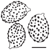
Spores of Cortinarius brunneocarpus (holotype). Scale bar 5 μm, del. H. Bashir.
Fig. 3
Fig. 3
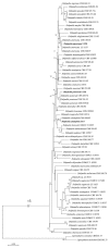
ITS phylogeny of sect. Hinnulei including the two new species and related taxa inferred from Bayesian inference analysis. New sequences are highlighted in boldface. BIPP ≥ 0.8 and MLBS ≥ 50 are indicated at the nodes.
Fig. 4
Fig. 4
Cortinarius lilacinoarmillatus (holotype). A. Basidiomata, B. Lamellar side of basidiomata. Photos K.C. Semwal.
Fig. 5
Fig. 5
Spores of Cortinarius lilacinoarmillatus (holotype). Scale bar 5 μm, del.V. Papp.
Fig. 6
Fig. 6
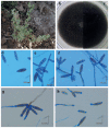
Curvularia khuzestanica (CBS 144736). a. The host plant, Atriplex lentiformis. b–c. Colony on PDA (front and reverse). d–f. Conidiophores and conidia. g–h. Mono- or bipolar germination of conidia.
Fig. 7
Fig. 7
ML phylogenetic tree reconstructed from a concatenated ITS–gpd dataset of 129 Curvularia strains representing previously described species and the her described C. khuzestanica. ML bootstrap values > 50 are shown at the nodes.
Fig. 8
Fig. 8
Gloeocantharellus neoechinosporus. 2–3. Basidiomata (a: GDGM75322; b: GDGM75321 Holotype!). 4–5. Basidiospores under SEM. 6. Basidiospores in Melzer’s reagent. Scale bars 2–3 50 mm, 4–6 5 μm.
Fig. 9
Fig. 9
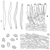
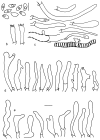
Gloeocantharellus neoechinosporus. 7. Gloeocystidia. 8. Basidia and gleocystidia. 9. Basidiospores. 10. Pileipellis. Scale bars 10 μm.
Fig. 10
Fig. 10
ML phylogenetic tree of Gloeocantharellus species reconstructed from ITS sequences. MLBS ≥ 50 and BIPP ≥ 0.9 are shown above or below the branches.
Fig. 11
Fig. 11
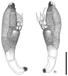
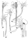
Laboulbenia bernaliana. Two mature thalli from slide FH00313719 (isotype). Scale bar 50 μm, del. A. De Kesel.
Fig. 12
Fig. 12
Laboulbenia oioveliicola. Two mature thalli from slide FH00313717 (isotype), and juvenile thallus from slide D. Haelew. 913d (holotype). Scale bar 100 μm, del. A. De Kesel.
Fig. 13
Fig. 13
Laboulbenia termiticola. a–d. Mature thalli from legs. e–f. Mature thallus from cephalon, with detail of perithecium and branching pattern of appendages. All thalli from slide BR5020195003490V (holotype). Scale bars a–e 100 μm, f 50 μm, del. A. De Kesel.
Fig. 14
Fig. 14
Basidiomata of Pluteus cutefractus, MCVE 30110 (holotype).
Fig. 15
Fig. 15
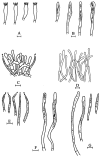
Pluteus cutefractus, MCVE 30110 (holotype). A, B. Cheilocystidia. C. Pleurocystidia. D. Pileipellis. E. Caulocystidia. F. Basidiospores. Scale bars 10 μm.
Fig. 16
Fig. 16
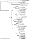
Maximum likelihood tree obtained from an ITS dataset of Pluteus sect. Celluloderma. MLBS ≥ 70 are presented above clade branches. Newly sequenced collections are highlighted in boldface.
Fig. 17
Fig. 17
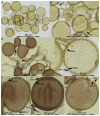
Rhizoglomus variabile. A–B. Spore clusters in PVLG with spores of very variable sizes. C. Spore cluster fragment in PVLG & Melzer’s reagent. Pigmented spores and subtending hyphae as well as the hyaline mycelia hyphae stain pinkish purple to purple. D. Spore in PVLG with three layers (SWL1, 2, 3) and a slightly funnel-shaped subtending hypha. E–G. Spores in PVLG & Melzer’s reagent. Hyaline outer and inner layers SWL1 and SWL3 do not stain in Melzer’s, while pigmented, structural layer SWL2 stains pinkish purple to purple. Subtending hypha typically cylindrical. Sometimes a septum can be recognized in some distance to the spore base, while the pore at the base regularly is open.
Fig. 18
Fig. 18
Phylogenetic tree obtained by analysis of partial SSU, ITS, and partial LSU rDNA sequences of different Rhizoglomus spp. Sequences labeled with GenBank accession numbers. Sequences obtained in this study shown in boldface. BI posterior probabilities ≥ 0.6 and ML bootstrap support ≥ 60 shown above and below branches, respectively. Thick branches represent clades ≥ 0.9/90 support.
Fig. 19
Fig. 19
Basidiomata and microscopic structures of Russula phloginea. a–b. Basidiomata, CNX530524068 (holotype). c. Basidiomata, CNX530524304. d. Sphaerocytes of lamellae trama in Congo Red reagent. e. Hymenium in Congo Red reagent. f. Hymenium in SV solution. g. Basidiospores in Melzer’s reagent. h–i. Basidiospores (SEM). Scale bars a, c 1 cm; b 1.5 cm; d–e, g–h 10 μm, f 20 μm, i 2 μm.
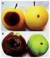
Fig. 20
Fig. 20
Microscopic structures of Russula phloginea, drawn from CNX530524068 (holotype). a. Basidia. b. Pleurocystidia. c. Terminal cell and pileocystidia in suprapellis. d. Pileocystidia in suprapellis. e. Pileocystidia in subpellis. f. Caulocystidia. Scale bars 10 μm.
Fig. 21
Fig. 21
ML phylogenetic tree of Russula subsect. Cyanoxanthinae reconstructed from the ITS–LSU–mtSSU–ef1α dataset. Branches are labeled with MLBS ≥ 50 and BIPP ≥ 0.95. New species shown in boldface.
Fig. 22
Fig. 22
Macro- and micro-morphology of Stagonosporopsis flacciduvarum. A. Colony morphology of UC23 isolate on MEA. B. Colony morphology on PDA. C. Colony morphology on OA. D. Pycnidia on PDA. E. Hyphae coil on PDA. F. Chlamydospore on PDA. G. Conidiogenous cells indicated by arrows on pine needle. H–I. Conidia on pine needle. J. Pycnidia on pine needle. K. Chlorotic area produced on berry infected by UC23 isolate. Scale bars D–F 50 μm, G–I 10 μm.
Fig. 23
Fig. 23
Results of pathogenicity assay in grapevine. Mycelial growth on berry and necrosis on young leaves after inoculum of different isolates at 25 °C after 7 d. A–C. Didymella americana UC30. D–F. D. calidophila CG7. G–I. D. pomorum UC56. J–L. Nothophoma quercina S3. M–O. Stagonosporopsis flacciduvarum UC23.
Fig. 24
Fig. 24
ML phylogenetic tree of Didymella species, reconstructed from the combined ITS–LSU–tub2rpb2 dataset. ML bootstrap support values shown at the nodes. Leptosphaeria conoidea and L. doliolum served as outgroups.
Fig. 25
Fig. 25
ML phylogenetic tree of Nothophoma species, reconstructed from the combined ITS–LSU–tub2rpb2 dataset. ML bootstrap support values shown at the nodes. Neopyrenochaeta telephoni served as outgroup.
Fig. 26
Fig. 26
ML phylogenetic tree of Stagonosporopsis species, reconstructed from the combined ITS–LSU–tub2rpb2 dataset. ML bootstrap support values shown at the nodes. Leptosphaeria conoidea served as outgroup.
Fig. 27
Fig. 27
Strobilomyces huangshanensis, HKAS 102613 (holotype). A. Basidiomata. B. Hymenophore. Photos: T. Guo. Scale bars = 1 cm.
Fig. 28
Fig. 28
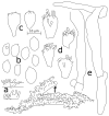
Microscopic characters of Strobilomyces huangshanensis, HKAS 102613 (holotype). A. Basidia and pleurocystidia. B. Cheilocystidia. C. Pileipellis. Scale bars 10 μm, del. L.H. Han.
Fig. 29
Fig. 29
Basidiospores of Strobilomyces huangshanensis under scanning electron microscope.
Fig. 30
Fig. 30
ML phylogenetic tree reconstructed from a four-locus dataset (rpb1, rpb2, tef1, and cox3). ML bootstrap support ≥ 50 and BI posterior probabilities ≥ 0.95 shown at the nodes. Strobilomyces huangshanensis sp. nov. shown in boldface.
Fig. 31
Fig. 31
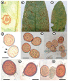
Uromyces klotzschianus, ISL-45963 (holotype). a. Abaxial view of infected Rumex dentatus leaf showing uredinial sorus characterized by two symptomatic rings. b. Abaxial view of leaf illustrating mixed uredinial and telial sori. c. Adaxial view showing mixed uredinial and telial sori. d–e. Urediniospores showing echinulate ornamentation (arrows). f. Urediniospores with teliospore indicating that they occur together. g–h. Teliospores with chocolate brown epispore and short hyaline pedicel. Scale bars d–f 40 μm, g–h 20 μm.
Fig. 32
Fig. 32
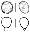
Uromyces klotzschianus. i–j. Echinulate urediniospores. k–l. Teliospores with smooth wall and short pedicel. Scale bars 20 μm, del. B. Ali.
Fig. 33
Fig. 33
ML phylogenetic reconstruction of the ITS dataset. Bootstrap values for 1000 replicates shown at nodes. New sequences highlighted in boldface.
Fig. 34
Fig. 34
Alternaria calendulae in India. a. Necrotic leaf spots followed by leaf tip and marginal blighting of Calendula officinalis caused by Alternaria calendulae. b. Spore morphology of A. calendulae. Scale bar = 50 μm. c. Development of leaf spot symptoms following artificial spray inoculation of A. calendulae on C. officinalis in greenhouse conditions.
Fig. 35
Fig. 35
Fruits with Alternaria rot symptoms. a. Quince (Cydonia oblonga) with black rot disease. b. Apple (Malus domestica, Golden delicious) with brown rot diseases.
Fig. 36
Fig. 36
Morphology of Alternaria tenuissima. a. After 7 d on PCA at 25 °C in 8 h fluorescens light/16 h dark cycle. b. Primary conidiophores. c. Conidial chain. d. Secondary conidiophore. Scale bars 10 μm.
Fig. 37
Fig. 37
Results of pathogenicity assay. Symptoms of Alternaria rot disease caused by inoculation with A. tenuissima (left fruits) and control treatments (right fruits) after 14 d at 25 °C with humidity above 90 %. a. Symptoms on apple. b. Symptoms on quince fruits.
Fig. 38
Fig. 38
ML phylogenetic tree of Alternaria isolates reconstructed from an ITS–gdp dataset. Bootstrap support values ≥ 50 indicated at the nodes.
Fig. 39
Fig. 39
Candelariella oleaginescens. a. Thallus with young apothecia. b. Thallus with apothecia. c. Ascoma in water, with, epihymenium, hymenium, and hypothecium. d. Ascospore in water. Scale bars a 250 μm, b 2 mm, c 100 μm, d 20 μm.
Fig. 40
Fig. 40
Rinodina sicula. a–b. Thallus with apothecia. c. Apothecium in water, with hymenium, ascus, and ascospores. d. Apothecium in water, with epihymenium, hymenium, ascus, ascospores. e. Ascus and ascospores in water. f. Ascospores in water. Scale bars a–b 2 mm, c 100 μm, d 50 μm, e–f 20 μm.
Fig. 41
Fig. 41
Verrucaria murina. a–b. Thallus with perithecia. c. Perithecium in water, with involucrellum, ascus, ascospores. d–f. Perithecium in water, with ascus and ascospores. Scale bars a–b 1 mm, c 50 μm, d–f 210 μm.
Fig. 42
Fig. 42
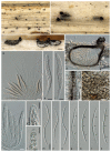
Lasiodiplodia theobromae on Dianella tasmanicavariegata’. A. Tip blight disease symptoms. B, C. Pycnidia on leaf surface. D. culture isolated from Dianella tasmanicavariegata’ on peptone salt agar (PSA). E. Pycnidium and conidia produced on PSA. F. Conidia after discharge from pycnidia. G. conidia after four weeks. Scale bars B 10 μm; C, E 100 μm; F–G 50 μm.
Fig. 43
Fig. 43
Marasmiellus subpruinosus BRNM 781138. Photo V. Antonín.
Fig. 44
Fig. 44
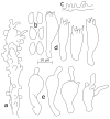
Marasmiellus subpruinosus BRNM 781138. a. Basidiospores. b. Basidia. c. Pileipellis elements. d. Cheilocystidia. e. Caulocystidia. Scale bar 10 μm, del. H. Ševčíková.
Fig. 45
Fig. 45
Phylogenetic placement of Marasmiellus subpruinosus among closely related species inferred from ITS rDNA sequences. Support values are given above the branches (≥ 0.90 for BI, ≥ 70 for ML).
Fig. 46
Fig. 46
Microstructures of Mycena albidolilacea. a. Caulocystidia. b. Basidiospores. c. Pileipellis hypha. d. Basidia. e. Cheilocystidia.
Fig. 47
Fig. 47
Microstructures of Mycena tenuispinosa. a. Pileipellis hyphae. b. Basidiospores. c. Basidia. d. Cheilocystidia. e. Caulocystidia. f. Apex of pileal “spinula”.
Fig. 48
Fig. 48
Microstructures of Mycena xantholeuca. a. Cheilocystidia. b. Pileipellis hyphae. c. Basidiospores. d. Basidia. e. Stipitipellis hyphae.
Fig. 49
Fig. 49
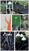
Basidiomata of studied Mycena species. a–d. Mycena tenuispinosa (LE 321754). e. Pileal “hair” of M. tenuispinosa at 400× (LE 321754). f–g. Mycena albidoloacea (f: LE 321756, g: LE 321757). h–i. Mycena xantholeuca (h: LE 321753, i: LE 321752).
Fig. 50
Fig. 50
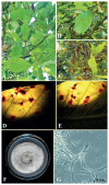
Neonectria neomacrospora. A. Initial small brown leaf spots on Madhuca longifolia. B. Characteristic yellow halos and reddish-brown zonation surrounding leaf spots. C. Marginal necroses of leaves. D. Surface view of leaf spots on M. longifolia leaf. E. Bottom view of leaf spots. F. Colony on PDA. G. Micro- and macroconidia. Scale bars D, E 1 cm; G 50 μm.
Fig. 51
Fig. 51
Plagiosphaera immersa (WU 40035). a, b. Translucent perithecia with laterally emerging ostioles immersed in dead stems of Urtica dioica. c, d. Vertical section of immersed perithecia showing the horizontally oriented perithecia with asci emerging from the lower half of the perithecial wall (d) and the apparently lateral (but in fact apical) ostioles bent upwards in more or less a right angle. e, f. Perithecial wall in section (e) and in face view (f). g. Hymenium with vital asci and paraphyses. h–i. Asci in vital state, detached from hymenium. j. Asci in dead state. k, l. Ascus apices with refractive apical ring. m–s. Vital (m–o) and dead (p–s) ascospores with 3 septa (indicated by arrows). All in water, except j, l, p–s in 3% KOH. Scale bars a 500 μm; b, c 100 μm; d 50 μm; e, f, h–j, m–s 10 μm; g 20 μm; k, l 5 μm.
Fig. 52
Fig. 52
ML phylogeny (-lnL = 28226.205) of selected Magnaporthales, reconstructed from the concatenated ITS–LSU–rpb1tef1 dataset, showing the phylogenetic position of Plagiosphaera immersa (in bold). ML/MP bootstrap support ≥ 70 are presented above or below the branches. Superscript T following taxon names indicates ex- epi-, holo-, or neotype isolates.
Fig. 53
Fig. 53
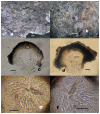
Macroscopic and microscopic morphology of Sphaerosporium lignatile (MICH 254984). A. Fruiting bodies are small, cushion-shaped masses of ochreous to honey-colored spores. B–D. Squash mounts in deionized water. B. We observed close associations between S. lignatile and bryophyte rhizoids. C. Hyphae are torulose, branched, and generate monoblastic conidia. D. Chlamydospore-like conidia, surrounded by a thick, hyaline, refractive cell wall. Scale bars A 1 mm, B–D 50 μm.
Fig. 54
Fig. 54
Ascomycota-wide phylogeny reconstructed from a six-locus data matrix (SSU, LSU, rpb1, rpb2, tef1, mitSSU). The topology is the result of ML inference performed with RAxML with all lineages collapsed to class level except for Pezizomycetes. For each node, MLBS ≥ 65 is presented above or below the branch leading to that node. Class Pezizomycetes and family Pyronemataceae highlighted by gray shading.

References

    1. Abbasi AM, Khan MA, Ahmad M, Zafar M. Medicinal plant biodiversity of lesser Himalayas-Pakistan. Springer Science & Business Media; Berlin: 2011.
    1. Accioly T, Sousa JO, Moreau P-A, Lécuru C, Silva BDB, Roy M, Gardes M, Baseia IG, Martín MP. Hidden fungal diversity from the Neotropics: Geastrum hirsutum, G. schweinitzii (Basidiomycota, Geastrales) and their allies. Plos One. 2019;14:e0211388. - PMC - PubMed
    1. Afshan N, Ishaq A, Niazi AR, Khalid AN. Geographical distribution and host range of genus Uromyces (Pucciniaceae, Uredinales) in Pakistan. International Conference on Chemical, Agricultural and Biological Sciences (CABS-2015); Istanbul (Turkey). 2015. pp. 48–54.
    1. Afshan NS, Khalid AN, Niazi AR, Iqbal SH. New records of Uredinales from Fairy Meadows, Pakistan. Mycotaxon. 2011;115:203–213.
    1. Ahmadpour SA, Mehrabi-Koushki M, Farokhinejad R. Neodidymelliopsis farokhinejadii, a new fungal species from dead branches of trees in Iran. Sydowia. 2017;69:171–182.

LinkOut - more resources