Treatment of Miller Class I Gingival Recession with Using Nonpedicle Adipose Tissue after Bichectomy Surgical Technique: A Case Report
- PMID: 31976089
- PMCID: PMC6955123
- DOI: 10.1155/2019/1049453
Treatment of Miller Class I Gingival Recession with Using Nonpedicle Adipose Tissue after Bichectomy Surgical Technique: A Case Report
Abstract
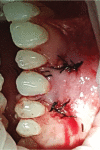
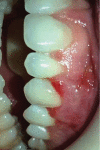
Gingival recession is an oral health problem that affects a large part of the population. Several treatments are suggested in the current literature; among them is the use of buccal fat pad grafting. The objective of this case report is to describe the treatment of a Miller Class I gingival recession using a nonpedicled buccal fat pad graft immediately after performing the surgery for buccal fat pad removal (bichectomy technique). First, bilateral surgical removal of the buccal fat pad was performed with the main objective of eliminating oral mucosa biting. The recipient site was prepared to receive a portion of the fat pad that was cut and macerated in a size that was sufficient to cover the recession. The patient was followed up at 15, 30, 60, and 365 days postsurgery, and the results showed an elimination of the oral mucosa biting and complete coverage of the gingival recession. It was concluded that the nonpedicled buccal fat pad graft is another option for the treatment of Miller Class I recessions.
Copyright © 2019 Carmen Lucia Mueller Storrer et al.
Conflict of interest statement
The authors declare that they have no conflicts of interest.
Figures


References
-
- Yared K. F. G., Zenobio E. G., Pacheco W. A etiologia multifatorial da recessão periodontal. Revista Dental Press de Ortodontia e Ortopedia Facial. 2006;11(6):45–51. doi: 10.1590/S1415-54192006000600007. - DOI
-
- Yadav A. P., Kulloli A., Shetty S., Ligade S. S., Martande S. S., Gholkar M. J. Sub-epithelial connective tissue graft for the management of Miller’s class I and class II isolated gingival recession defect: a systematic review of the factors influencing the outcome. Journal of Investigative and Clinical Dentistry. 2018;9(3, article e12325) doi: 10.1111/jicd.12325. - DOI - PubMed
Publication types
LinkOut - more resources
Full Text Sources

