

Normal Anatomy and Anomalies of the Rectus Extraocular Muscles in Human: A Review of the Recent Data and Findings
- PMID: 31976329
- PMCID: PMC6954479
- DOI: 10.1155/2019/8909162
Normal Anatomy and Anomalies of the Rectus Extraocular Muscles in Human: A Review of the Recent Data and Findings
Abstract
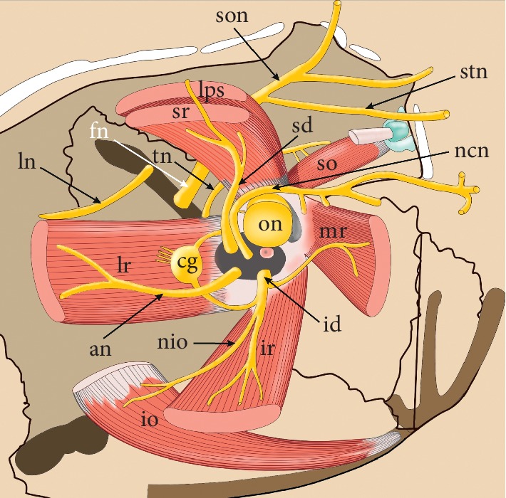
Development of modern surgical techniques is associated with the need for a thorough knowledge of surgical anatomy and, in the case of ophthalmologic surgery, also functional aspects of extraocular muscles. Thus, the leading idea of this review was to summarize the most recent findings regarding the normal anatomy and anomalies of the extraocular rectus muscles (ERMs). Particular attention was paid to the presentation of detailed and structured data on the gross anatomy of the ERMs, including their attachments, anatomical relationships, vascularization, and innervation. This issue of ERMs innervation was presented in detail, considering the research that has recently been carried out on human material using advanced anatomical techniques such as Sihler's technique of the nerves staining. The text was supplemented with a carefully selected graphic material (including anatomical specimens prepared specially for the purpose of this review) and discussion of the clinical cases and practical significance of the presented issues.
Copyright © 2019 Robert Haładaj.
Conflict of interest statement
The authors have no conflicts of interest to declare.
Figures


Similar articles
-
Branching patterns of rabbit oculomotor and trochlear nerves demonstrated by Sihler's stain technique.Biotech Histochem. 2002 Jan;77(1):21-5. Biotech Histochem. 2002. PMID: 11991327
-
Comparison of lateral and medial rectus muscle in human: an anatomical study with particular emphasis on morphology, intramuscular innervation pattern variations and discussion on clinical significance.Surg Radiol Anat. 2020 May;42(5):607-616. doi: 10.1007/s00276-019-02400-x. Epub 2020 Jan 2. Surg Radiol Anat. 2020. PMID: 31897658
-
Comparison of the Superior and Inferior Rectus Muscles in Humans: An Anatomical Study with Notes on Morphology, Anatomical Variations, and Intramuscular Innervation Patterns.Biomed Res Int. 2020 Apr 30;2020:9037693. doi: 10.1155/2020/9037693. eCollection 2020. Biomed Res Int. 2020. PMID: 32420380 Free PMC article.
-
The anatomy and physiology of the ocular motor system.Handb Clin Neurol. 2011;102:21-69. doi: 10.1016/B978-0-444-52903-9.00008-X. Handb Clin Neurol. 2011. PMID: 21601062 Review.
-
Sihler's whole mount nerve staining technique: a review.Biotech Histochem. 2010 Feb;85(1):19-42. doi: 10.3109/10520290903048384. Biotech Histochem. 2010. PMID: 19572223 Free PMC article. Review.
Cited by
-
Variations in the anterior thoracic wall with sternalis muscle and accessory pectoralis major muscle.Surg Radiol Anat. 2022 May;44(5):785-790. doi: 10.1007/s00276-022-02923-w. Epub 2022 Mar 27. Surg Radiol Anat. 2022. PMID: 35344059
-
Pharmacologically Induced Accommodation Palsy and the Bioelectrical Activity of the Muscular System: A Preliminary Investigation.Diagnostics (Basel). 2024 May 4;14(9):961. doi: 10.3390/diagnostics14090961. Diagnostics (Basel). 2024. PMID: 38732375 Free PMC article.
-
Clinical Impact of Specific Extraocular Muscle Manipulation and the Oculocardiac Reflex on Postoperative Vomiting in Pediatric Strabismus Surgery: A Multicenter, Observational Study.Paediatr Anaesth. 2025 Feb;35(2):163-174. doi: 10.1111/pan.15047. Epub 2024 Nov 30. Paediatr Anaesth. 2025. PMID: 39614704 Free PMC article.
-
Relationship between Axial Length and Corneo-Scleral Topography: A Preliminary Study.Diagnostics (Basel). 2021 Mar 18;11(3):542. doi: 10.3390/diagnostics11030542. Diagnostics (Basel). 2021. PMID: 33803709 Free PMC article.
-
Topographical anatomy of the annulus of Zinn.Sci Rep. 2022 Jan 20;12(1):1064. doi: 10.1038/s41598-022-05178-y. Sci Rep. 2022. PMID: 35058545 Free PMC article.
References
-
- Farzavandi S. Surgical anatomy. In: Farzavandi S., editor. Color Atlas of Strabismus Surgery. New York, NY, USA: Springer; 2007. pp. 91–101.
Publication types
MeSH terms
Substances
LinkOut - more resources
Full Text Sources
