DMD carrier model with mosaic dystrophin expression in the heart reveals complex vulnerability to myocardial injury
- PMID: 31976522
- PMCID: PMC7158376
- DOI: 10.1093/hmg/ddaa015
DMD carrier model with mosaic dystrophin expression in the heart reveals complex vulnerability to myocardial injury
Abstract
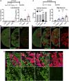
Duchenne muscular dystrophy (DMD) is a devastating neuromuscular disease that causes progressive muscle wasting and cardiomyopathy. This X-linked disease results from mutations of the DMD allele on the X-chromosome resulting in the loss of expression of the protein dystrophin. Dystrophin loss causes cellular dysfunction that drives the loss of healthy skeletal muscle and cardiomyocytes. As gene therapy strategies strive toward dystrophin restoration through micro-dystrophin delivery or exon skipping, preclinical models have shown that incomplete restoration in the heart results in heterogeneous dystrophin expression throughout the myocardium. This outcome prompts the question of how much dystrophin restoration is sufficient to rescue the heart from DMD-related pathology. Female DMD carrier hearts can shed light on this question, due to their mosaic cardiac dystrophin expression resulting from random X-inactivation. In this work, a dystrophinopathy carrier mouse model was derived by breeding male or female dystrophin-null mdx mice with a wild type mate. We report that these carrier hearts are significantly susceptible to injury induced by one or multiple high doses of isoproterenol, despite expressing ~57% dystrophin. Importantly, only carrier mice with dystrophic mothers showed mortality after isoproterenol. These findings indicate that dystrophin restoration in approximately half of the heart still allows for marked vulnerability to injury. Additionally, the discovery of divergent stress-induced mortality based on parental origin in mice with equivalent dystrophin expression underscores the need for better understanding of the epigenetic, developmental, and even environmental factors that may modulate vulnerability in the dystrophic heart.
© The Author(s) 2020. Published by Oxford University Press. All rights reserved. For Permissions, please email: journals.permissions@oup.com.
Figures


Similar articles
-
Exclusive skeletal muscle correction does not modulate dystrophic heart disease in the aged mdx model of Duchenne cardiomyopathy.Hum Mol Genet. 2013 Jul 1;22(13):2634-41. doi: 10.1093/hmg/ddt112. Epub 2013 Mar 3. Hum Mol Genet. 2013. PMID: 23459935 Free PMC article.
-
Prevention of exercised induced cardiomyopathy following Pip-PMO treatment in dystrophic mdx mice.Sci Rep. 2015 Mar 11;5:8986. doi: 10.1038/srep08986. Sci Rep. 2015. PMID: 25758104 Free PMC article.
-
Enhanced dimethylarginine degradation improves coronary flow reserve and exercise tolerance in Duchenne muscular dystrophy carrier mice.Am J Physiol Heart Circ Physiol. 2020 Sep 1;319(3):H582-H603. doi: 10.1152/ajpheart.00333.2019. Epub 2020 Aug 7. Am J Physiol Heart Circ Physiol. 2020. PMID: 32762558 Free PMC article.
-
Molecular correction of Duchenne muscular dystrophy by splice modulation and gene editing.RNA Biol. 2021 Jul;18(7):1048-1062. doi: 10.1080/15476286.2021.1874161. Epub 2021 Jan 20. RNA Biol. 2021. PMID: 33472516 Free PMC article. Review.
-
The multifaceted view of heart problem in Duchenne muscular dystrophy.Cell Mol Life Sci. 2021 Jul;78(14):5447-5468. doi: 10.1007/s00018-021-03862-2. Epub 2021 Jun 6. Cell Mol Life Sci. 2021. PMID: 34091693 Free PMC article. Review.
Cited by
-
Cardiac Involvement in Dystrophin-Deficient Females: Current Understanding and Implications for the Treatment of Dystrophinopathies.Genes (Basel). 2020 Jul 8;11(7):765. doi: 10.3390/genes11070765. Genes (Basel). 2020. PMID: 32650403 Free PMC article. Review.
-
Impact of estrogen deficiency on diaphragm and leg muscle contractile function in female mdx mice.PLoS One. 2021 Mar 31;16(3):e0249472. doi: 10.1371/journal.pone.0249472. eCollection 2021. PLoS One. 2021. PMID: 33788896 Free PMC article.
-
Functional cardiac consequences of β-adrenergic stress-induced injury in a model of Duchenne muscular dystrophy.Dis Model Mech. 2024 Oct 1;17(10):dmm050852. doi: 10.1242/dmm.050852. Epub 2024 Oct 9. Dis Model Mech. 2024. PMID: 39268580 Free PMC article.
-
Functional cardiac consequences of β-adrenergic stress-induced injury in the mdx mouse model of Duchenne muscular dystrophy.bioRxiv [Preprint]. 2024 Apr 20:2024.04.15.589650. doi: 10.1101/2024.04.15.589650. bioRxiv. 2024. Update in: Dis Model Mech. 2024 Oct 1;17(10):dmm050852. doi: 10.1242/dmm.050852. PMID: 38659739 Free PMC article. Updated. Preprint.
References
-
- Nigro G., Comi L.I., Politano L. and Bain R.J.I. (1990) The incidence and evolution of cardiomyopathy in Duchenne muscular dystrophy. Int. J. Cardiol., 26, 271–277. - PubMed
-
- Birnkrant D.J., Bushby K., Bann C.M., Alman B.A., Apkon S.D., Blackwell A., Case L.E., Cripe L.H., Hadjiyannakis S., Olson A.K. et al. (2018) Diagnosis and management of Duchenne muscular dystrophy, part 2: respiratory, cardiac, bone health, and orthopaedic management. Lancet Neurol., 17, 347–361. - PMC - PubMed
-
- Spurney C.F. (2011) Cardiomyopathy of Duchenne muscular dystrophy: current understanding and future directions. Muscle Nerve, 44, 8–19. - PubMed
-
- Muntoni F. (2003) Cardiomyopathy in muscular dystrophies. Curr. Opin. Neurol., 16, 577–583. - PubMed
Publication types
MeSH terms
Substances
Grants and funding
LinkOut - more resources
Full Text Sources
Medical
Research Materials

