Multicenter Study
. 2020 Nov 1;116(13):2116-2130.
doi: 10.1093/cvr/cvaa010.

Impact of functional studies on exome sequence variant interpretation in early-onset cardiac conduction system diseases

Affiliations
Multicenter Study

Impact of functional studies on exome sequence variant interpretation in early-onset cardiac conduction system diseases

Kenshi Hayashi et al. Cardiovasc Res. .

Abstract

Aims: The genetic cause of cardiac conduction system disease (CCSD) has not been fully elucidated. Whole-exome sequencing (WES) can detect various genetic variants; however, the identification of pathogenic variants remains a challenge. We aimed to identify pathogenic or likely pathogenic variants in CCSD patients by using WES and 2015 American College of Medical Genetics and Genomics (ACMG) standards and guidelines as well as evaluating the usefulness of functional studies for determining them.

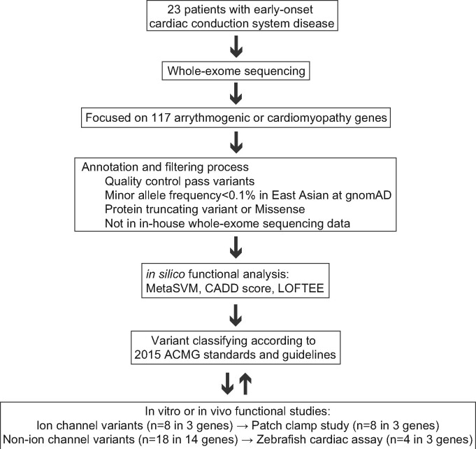
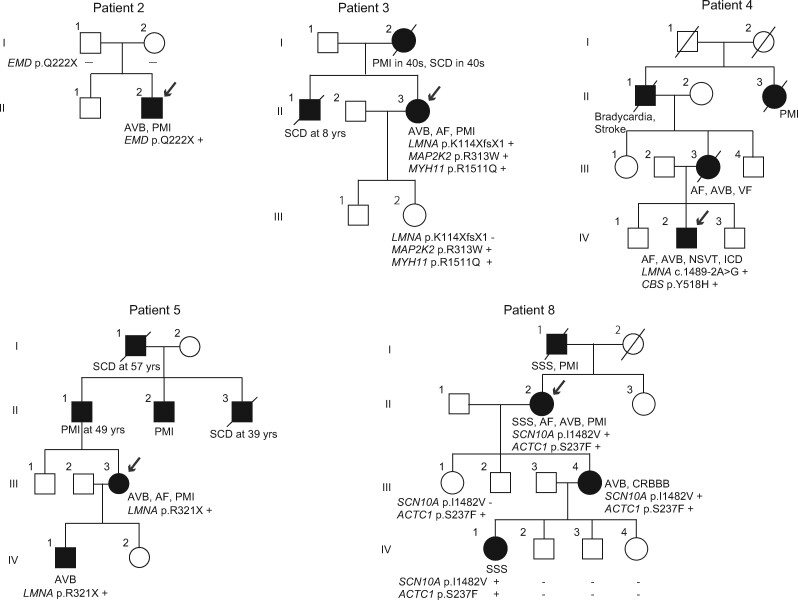
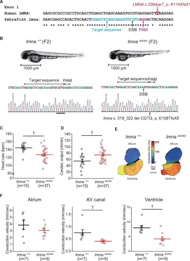
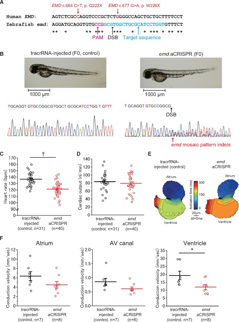
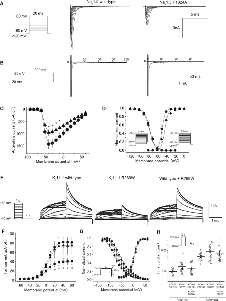
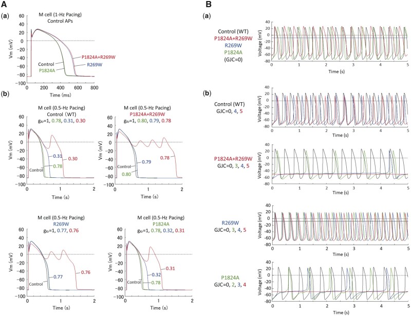
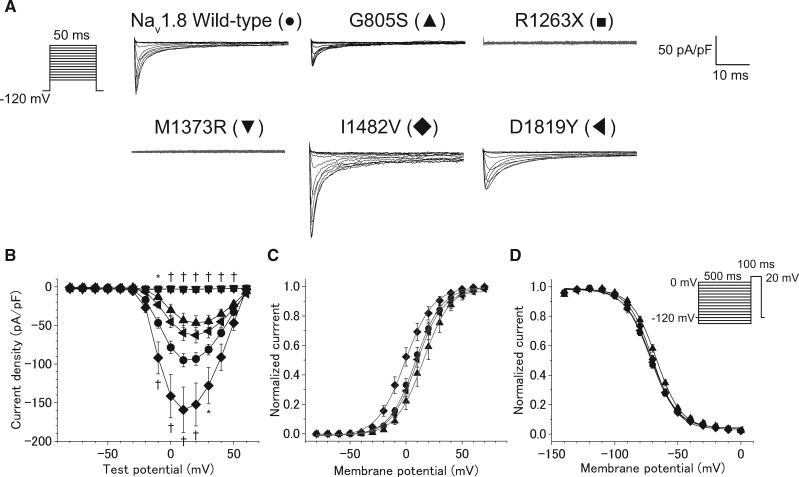
Methods and results: We performed WES of 23 probands diagnosed with early-onset (<65 years) CCSD and analysed 117 genes linked to arrhythmogenic diseases or cardiomyopathies. We focused on rare variants (minor allele frequency < 0.1%) that were absent from population databases. Five probands had protein truncating variants in EMD and LMNA which were classified as 'pathogenic' by 2015 ACMG standards and guidelines. To evaluate the functional changes brought about by these variants, we generated a knock-out zebrafish with CRISPR-mediated insertions or deletions of the EMD or LMNA homologs in zebrafish. The mean heart rate and conduction velocities in the CRISPR/Cas9-injected embryos and F2 generation embryos with homozygous deletions were significantly decreased. Twenty-one variants of uncertain significance were identified in 11 probands. Cellular electrophysiological study and in vivo zebrafish cardiac assay showed that two variants in KCNH2 and SCN5A, four variants in SCN10A, and one variant in MYH6 damaged each gene, which resulted in the change of the clinical significance of them from 'Uncertain significance' to 'Likely pathogenic' in six probands.

Conclusion: Of 23 CCSD probands, we successfully identified pathogenic or likely pathogenic variants in 11 probands (48%). Functional analyses of a cellular electrophysiological study and in vivo zebrafish cardiac assay might be useful for determining the pathogenicity of rare variants in patients with CCSD. SCN10A may be one of the major genes responsible for CCSD.

Keywords: CRISPR/Cas9-mediated gene knock-out in zebrafish; 2015 ACMG standards and guidelines; Cardiac conduction system disease; Cellular electrophysiological study; Whole exome sequencing.


Figures

Figure 1
Figure 1
Experimental workflow for determining pathogenicity of candidate variants in CCSD by targeted analysis of WES. ACMG, American College of Medical Genetics and Genomics; CADD, combined annotation dependent depletion; gnomAD, Genome Aggregation Database; LOFTEE, loss-of-function transcript effect estimator; MetaSVM, in silico ensemble damaging score.
Figure 2
Figure 2
Pedigrees of family 2, 3, 4, 5, and 8. Squares and circles represent males and females, respectively. Slash marks represent deceased individuals. Black filled symbols indicate patients with a clinical diagnosis of CCSD. Open symbols represent unaffected family members. The probands are indicated by arrows. Plus and minus signs indicate positive and negative variants, respectively.
Figure 3
Figure 3
Functional studies of LMNA c.339dupT using F2 zebrafish embryos with lmna deletion mutation. (A) Multiple sequence alignment of human LMNA and zebrafish lmna. DSB, double-strand break; PAM, protospacer adjacent motif. (B) Representative images illustrating the morphology of 2 dpf lmna+/+ (wild-type) and lmnadel/del mutants, and Sanger sequence of lmna gene. (C) HR of lmna+/+ (n = 15) and lmnadel/del mutants (n = 37). (D) Cardiac output of lmna+/+ (n = 15) and lmnadel/del mutants (n = 37). (E) Isochronal map of lmna+/+ and lmnadel/del mutants summarizing the regional spread of electrical activity across the atrium and into the ventricle. The lines represent the positions of the action potential wavefront at 5-ms intervals. The colour scale depicts the timing of electrical activation (blue areas activated before red areas). (F) Mean estimated conduction velocities at the atrium, AV, atrioventricular canal, and ventricle of lmna+/+ (n = 7) and lmnadel/del mutants (n = 9). Regions of interest was placed at middle of atrium, AV canal, or ventricle. P < 0.01.
Figure 4
Figure 4
Functional studies on EMD p. Q222X and p. W226X using CRISPR-mediated deletions of the human EMD ortholog, emd, in zebrafish. (A) Multiple sequence alignment of human EMD and zebrafish emd. (B) Representative images illustrating the morphology of 2 dpf emd aCRISPR and tracrRNA-injected embryos and Sanger sequence of emd gene. (C) HR of tracrRNA-injected (n = 31) and emd aCRISPR (n = 40) embryos. (D) Cardiac output of tracrRNA-injected (n = 31) and emd aCRISPR (n = 40) embryos. (E) Isochronal map of tracrRNA-injected and emd aCRISPR embryos summarizing the regional spread of electrical activity across the atrium and into the ventricle. (F) Mean estimated conduction velocities at the atrium, AV canal, and ventricle of tracrRNA-injected (n = 7) and emd aCRISPR (n = 8) embryos. Regions of interest was placed at the middle of the atrium, AV canal, or ventricle. P < 0.01; *P < 0.05.
Figure 5
Figure 5
Functional properties of Nav1.5 channel and Kv11.1 channel in a patient with SCN5A P1824A and KCNH2 R269W. (A) The voltage protocol and representative whole-cell Na+ currents of the wild-type and P1824A Nav1.5 channels. (B) Comparison of late Na+ currents. The late Na+ currents were measured at the end of 200-ms depolarizing pulses, as shown in the inset. (C) I–V relationships for peak currents in HEK293 cells transfected with wild-type (closed circle, n = 23) and P1824A (closed triangle, n = 21). *P < 0.05 vs. wild-type. (D) The voltage protocols and the voltage dependence of steady-state fast inactivation and activation for wild-type (n = 19 and 23) and P1824A (n = 14 and 21). (E) The voltage protocol and representative expressed currents in CHO-K1 cells transfected with Kv11.1 wild-type alone, Kv11.1 R269W, and wild-type plus R269W. (F) I–V relationships for tail currents in CHO-K1 cells transfected with wild-type alone (closed circle, n = 19), R269W (closed square, n = 17), and wild-type plus R269W (closed triangle, n = 12). *P < 0.05 vs. wild-type. (G) The voltage protocols and normalized steady-state activation and inactivation curves for wild-type alone (n = 19 and 7), R269W (n = 17 and 10), and wild-type plus R269W (n = 12 and 9). (H) The voltage protocol and fast and slow components of deactivation time constants as a function of test potentials for wild-type alone (n = 15), R269W (n = 18), and wild-type plus R269W (n = 20). The deactivation process was fit to biexponential functions.
Figure 6
Figure 6
Computer simulation of the effects of modified kinetic behaviour of Kv11.1 and Nav1.5 currents on electrophysiological properties of human ventricular myocytes (mid-myocardial cells) and electrotonic effects of atrial myocytes on pacemaker activity of peripheral sinoatrial node (SAN) cells in a normal subject and a patient with modified Kv11.1 and Nav1.5 currents. (A) Simulated action potentials of ventricular mid-myocardial cell models for a normal subject and patient with modified Kv11.1 and Nav1.5 currents (Aa). The model cells were paced at 1 Hz by 1-ms stimuli of 60 pA/pF for 30 min; steady-state behaviours after the last stimulus are shown. Modified inactivation/deactivation kinetics of R269W (KCNH2 R269W), P1824A (SCN5A P1824A), and P1824A+R269W (KCNH2 R269W and SCN5A P1824A) prolonged action potential duration at 90% repolarization (APD90) from 400 to 517, 404, or 526 ms, respectively. Effects of IKr block on action potentials were also determined for the control and patient models, and those with KCNH2 or SCN5A single mutation (Ab). (B) Simulated spontaneous action potentials in peripheral SAN cell models for the control, patient, and KCNH2 or SCN5A single mutation (Ba). Action potentials were also computed for each SAN model cell connected to an atrial cell with gap junction conductance (GJC) of 0–5 nS (Bb), as described previously.
Figure 7
Figure 7
The functional consequence of five variants in SCN10A assessed by whole-cell patch clamp recording. (A) The voltage protocol and representative current traces of Nav 1.8 using wild-type and mutant channels. (B) I–V relationships for peak currents in ND 7/23 cells transfected with SCN10A wild-type (●, n = 64) and five variants including G805S (▲, n = 15), R1263X (■, n = 15), M1373R (▼, n = 19), I1482V (◆, n = 25), and D1819Y (◀, n = 24). *P < 0.05 or P < 0.01 vs. wild-type by one-way ANOVA, followed by a Bonferroni post hoc test. (C) Normalized steady-state activation curves of SCN10A wild-type (n = 64), and three variants including G805S (n = 15), I1482V (n = 25), and D1819Y (n = 24). (D) The voltage protocols and normalized steady-state inactivation curves of SCN10A wild-type (n = 33) and three variants including G805S (n = 13), I1482V (n = 23), and D1819Y (n = 22).

References

    1. Beinart R, Ruskin J, Milan D. The genetics of conduction disease. Heart Fail Clin 2010;6:201–214. - PubMed
    1. Wolf CM, Berul CI. Inherited conduction system abnormalities–one group of diseases, many genes. J Cardiovasc Electrophysiol 2006;17:446–455. - PubMed
    1. Ishikawa T, Tsuji Y, Makita N. Inherited bradyarrhythmia: a diverse genetic background. J Arrhythm 2016;32:352–358. - PMC - PubMed
    1. Celestino-Soper PB, Doytchinova A, Steiner HA, Uradu A, Lynnes TC, Groh WJ, Miller JM, Lin H, Gao H, Wang Z, Liu Y, Chen PS, Vatta M. Evaluation of the genetic basis of familial aggregation of pacemaker implantation by a large next generation sequencing panel. PLoS One 2015;10:e0143588. - PMC - PubMed
    1. Ho CY, Charron P, Richard P, Girolami F, Van Spaendonck-Zwarts KY, Pinto Y. Genetic advances in sarcomeric cardiomyopathies: state of the art. Cardiovasc Res 2015;105:397–408. - PMC - PubMed

Publication types

MeSH terms