

Intermittent Starvation Extends the Functional Lifetime of Primary Human Hepatocyte Cultures
- PMID: 31977024
- PMCID: PMC7098377
- DOI: 10.1093/toxsci/kfaa003
Intermittent Starvation Extends the Functional Lifetime of Primary Human Hepatocyte Cultures
Abstract
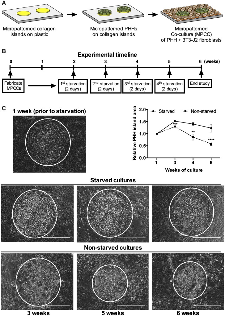
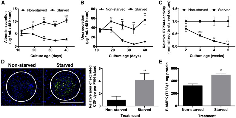
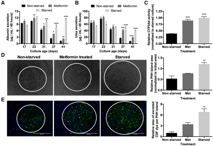
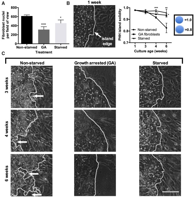
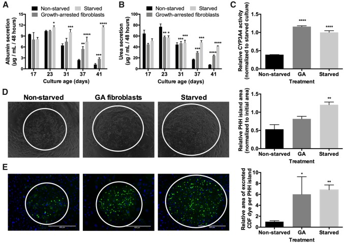
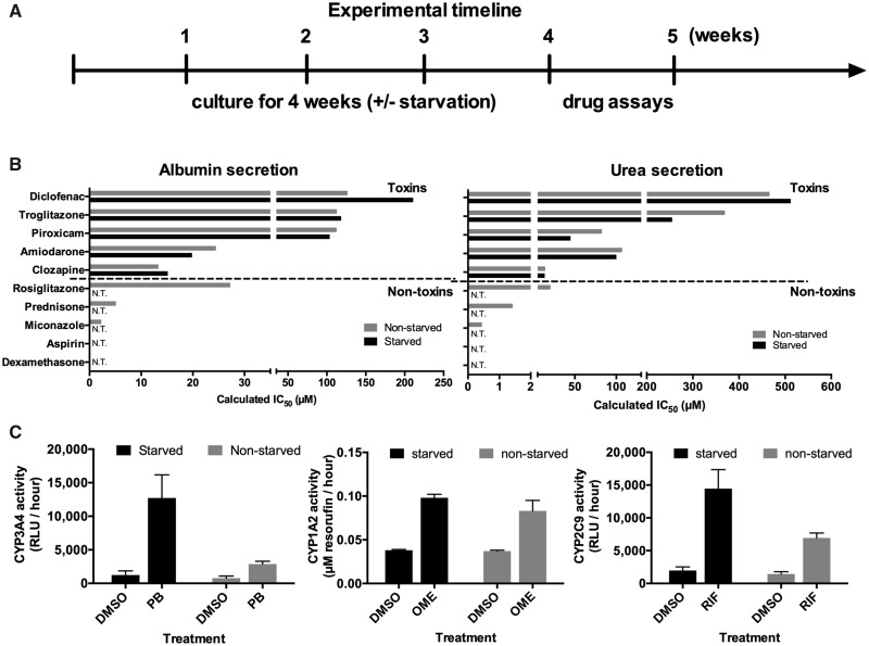
Primary human hepatocyte (PHH) cultures have become indispensable to mitigate the risk of adverse drug reactions in human patients. In contrast to dedifferentiating monocultures, coculture with nonparenchymal cells maintains PHH functions for 2-4 weeks. However, because the functional lifespan of PHHs in vivo is 200-400 days, it is desirable to further prolong PHH functions in vitro toward modeling chronic drug exposure and disease progression. Fasting has benefits on the longevity of organisms and the health of tissues such as the liver. We hypothesized that a culturing protocol that mimics dynamic fasting/starvation could activate starvation pathways and prolong PHH functional lifetime. To mimic starvation, serum and hormones were intermittently removed from the culture medium of micropatterned cocultures (MPCCs) containing PHHs organized onto collagen domains and surrounded by 3T3-J2 murine fibroblasts. A weekly 2-day starvation optimally prolonged PHH functional lifetime for 6+ weeks in MPCCs versus a decline after 3 weeks in nonstarved controls. The 2-day starvation also enhanced the functions of PHH monocultures for 2 weeks, suggesting direct effects on PHHs. In MPCCs, starvation activated 5' adenosine monophosphate-activated protein kinase (AMPK) and restricted fibroblast overgrowth onto PHH islands, thereby maintaining hepatic polarity. The effects of starvation on MPCCs were partially recapitulated by activating AMPK using metformin or growth arresting fibroblasts via mitomycin-C. Lastly, starved MPCCs demonstrated lower false positives for drug toxicity tests and higher drug-induced cytochrome-P450 activities versus nonstarved controls even after 5 weeks. In conclusion, intermittent serum/hormone starvation extends PHH functional lifetime toward enabling clinically relevant drug screening.
Keywords: AMPK; fasting; metformin; micropatterned coculture.
© The Author(s) 2020. Published by Oxford University Press on behalf of the Society of Toxicology. All rights reserved. For permissions, please e-mail: journals.permissions@oup.com.
Figures
Similar articles
-
Physiologically inspired culture medium prolongs the lifetime and insulin sensitivity of human hepatocytes in micropatterned co-cultures.Toxicology. 2021 Feb 15;449:152662. doi: 10.1016/j.tox.2020.152662. Epub 2020 Dec 24. Toxicology. 2021. PMID: 33359713 Free PMC article.
-
Microengineered cultures containing human hepatic stellate cells and hepatocytes for drug development.Integr Biol (Camb). 2017 Aug 14;9(8):662-677. doi: 10.1039/c7ib00027h. Integr Biol (Camb). 2017. PMID: 28702667
-
Micropatterned Coculture With 3T3-J2 Fibroblasts Enhances Hepatic Functions and Drug Screening Utility of HepaRG Cells.Toxicol Sci. 2021 Apr 27;181(1):90-104. doi: 10.1093/toxsci/kfab018. Toxicol Sci. 2021. PMID: 33590212
-
[Application of Human Liver Organoids for Pharmaceutical Research].Yakugaku Zasshi. 2025;145(3):189-194. doi: 10.1248/yakushi.24-00177-4. Yakugaku Zasshi. 2025. PMID: 40024731 Review. Japanese.
-
Bioengineered Liver Models for Drug Testing and Cell Differentiation Studies.Cell Mol Gastroenterol Hepatol. 2017 Dec 6;5(3):426-439.e1. doi: 10.1016/j.jcmgh.2017.11.012. eCollection 2018 Mar. Cell Mol Gastroenterol Hepatol. 2017. PMID: 29675458 Free PMC article. Review.
Cited by
-
Physiologically inspired culture medium prolongs the lifetime and insulin sensitivity of human hepatocytes in micropatterned co-cultures.Toxicology. 2021 Feb 15;449:152662. doi: 10.1016/j.tox.2020.152662. Epub 2020 Dec 24. Toxicology. 2021. PMID: 33359713 Free PMC article.
-
Effects of Fasting on THP1 Macrophage Metabolism and Inflammatory Profile.Int J Mol Sci. 2024 Aug 20;25(16):9029. doi: 10.3390/ijms25169029. Int J Mol Sci. 2024. PMID: 39201723 Free PMC article.
-
Metformin Protects ARPE-19 Cells from Glyoxal-Induced Oxidative Stress.Oxid Med Cell Longev. 2020 Jul 9;2020:1740943. doi: 10.1155/2020/1740943. eCollection 2020. Oxid Med Cell Longev. 2020. PMID: 32695253 Free PMC article.
-
Assessing the compatibility of primary human hepatocyte culture within porous silk sponges.RSC Adv. 2020 Oct 12;10(62):37662-37674. doi: 10.1039/d0ra04954a. eCollection 2020 Oct 12. RSC Adv. 2020. PMID: 35515172 Free PMC article.
-
Challenges for the Applications of Human Pluripotent Stem Cell-Derived Liver Organoids.Front Cell Dev Biol. 2021 Oct 1;9:748576. doi: 10.3389/fcell.2021.748576. eCollection 2021. Front Cell Dev Biol. 2021. PMID: 34660606 Free PMC article. Review.
References
-
- Abboud G., Kaplowitz N. (2007). Drug-induced liver injury. Drug Saf. 30, 277–294. - PubMed
-
- Berger D. R., Ware B. R., Davidson M. D., Allsup S. R., Khetani S. R. (2015). Enhancing the functional maturity of induced pluripotent stem cell-derived human hepatocytes by controlled presentation of cell-cell interactions in vitro. Hepatology 61, 1370–1381. - PubMed
Publication types
MeSH terms
Substances
Grants and funding
LinkOut - more resources
Full Text Sources
