. 2020 Mar;13(3):e008303.
doi: 10.1161/CIRCEP.119.008303. Epub 2020 Jan 24.

Pulsed Field Ablation Versus Radiofrequency Ablation: Esophageal Injury in a Novel Porcine Model

Affiliations

Pulsed Field Ablation Versus Radiofrequency Ablation: Esophageal Injury in a Novel Porcine Model

Jacob S Koruth et al. Circ Arrhythm Electrophysiol. 2020 Mar.

Abstract

Background: Pulsed field ablation (PFA) can be myocardium selective, potentially sparing the esophagus during left atrial ablation. In an in vivo porcine esophageal injury model, we compared the effects of newer biphasic PFA with radiofrequency ablation (RFA).

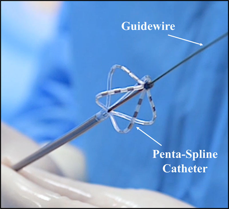
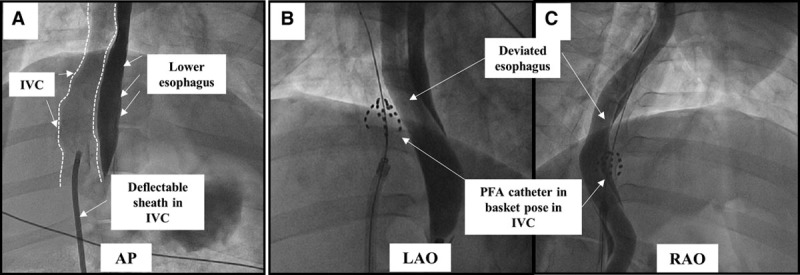
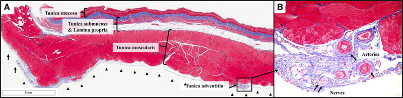
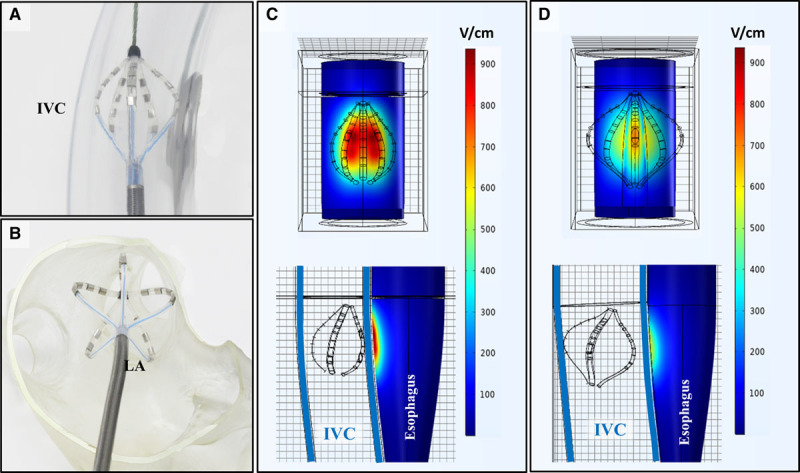
Methods: In 10 animals, under general anesthesia, the lower esophagus was deflected toward the inferior vena cava using an esophageal deviation balloon, and ablation was performed from within the inferior vena cava at areas of esophageal contact. Four discrete esophageal sites were targeted in each animal: 6 animals received 8 PFA applications/site (2 kV, multispline catheter), and 4 animals received 6 clusters of irrigated RFA applications (30 W×30 seconds, 3.5 mm catheter). All animals were survived to 25 days, sacrificed, and the esophagus submitted for pathological examination, including 10 discrete histological sections/esophagus.

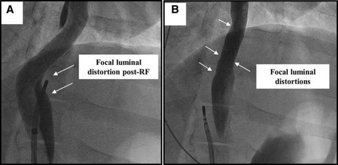
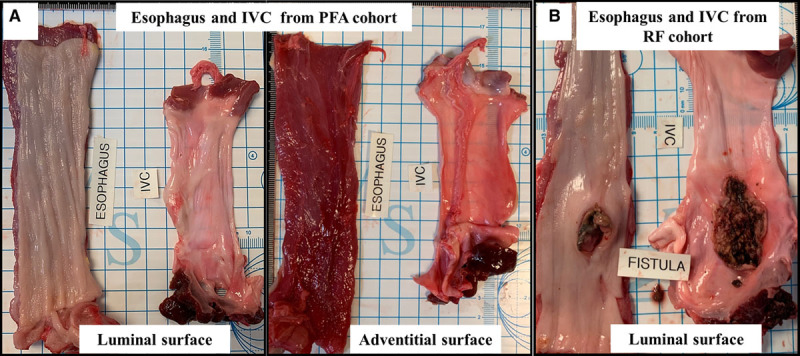
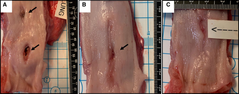
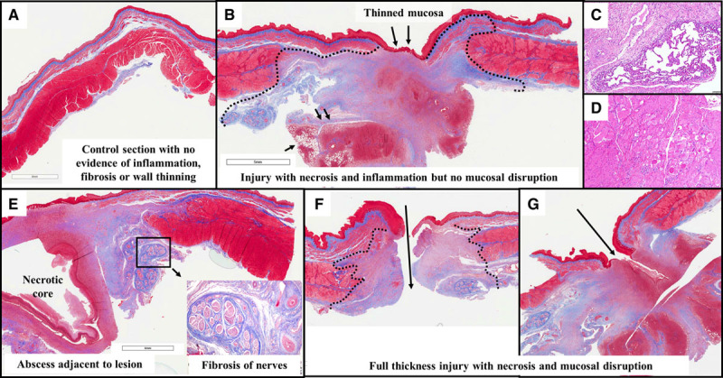
Results: The animals weight increased by 13.7±6.2% and 6.8±6.3% (P=0.343) in the PFA and RFA cohorts, respectively. No PFA animals (0 of 6, 0%) developed abnormal in-life observations, but 1 of 4 RFA animals (25%) developed fever and dyspnea. On necropsy, no PFA animals (0 of 6, 0%) demonstrated esophageal lesions. In contrast, esophageal injury occurred in all RFA animals (4 of 4, 100%; P=0.005): a mean of 1.5 mucosal lesions/animal (length, -21.8±8.9 mm; width, -4.9±1.4 mm) were observed, including one esophago-pulmonary fistula and deep esophageal ulcers in the other animals. Histological examination demonstrated tissue necrosis surrounded by acute and chronic inflammation and fibrosis. The necrotic RFA lesions involved multiple esophageal tissue layers with evidence of arteriolar medial thickening and fibrosis of periesophageal nerves. Abscess formation and full-thickness esophageal wall disruptions were seen in areas of perforation/fistula.

Conclusions: In this novel porcine model of esophageal injury, biphasic PFA induced no chronic histopathologic esophageal changes, while RFA demonstrated a spectrum of esophageal lesions including fistula and deep esophageal ulcers and abscesses.

Keywords: atrial fibrillation; catheter ablation; electroporation; fistula; swine.


Figures

Figure 1.
Figure 1.
Pulsed field ablation (PFA) catheter: pentaspline over-the-wire PFA catheter in basket pose.
Figure 2.
Figure 2.
Fluoroscopic view of the esophageal injury mode: pulsed field ablation (PFA) cohort. A, Contrast angiography was performed using a long deflectable sheath placed in the inferior vena cava (IVC; outlined). In the anteroposterior (AP) view, the IVC is seen rightward of the contrast filled esophagus. B and C, Left and right anterior oblique (LAO and RAO) projections demonstrate the PFA catheter in basket pose forcefully pushed against the deviated esophagus. The PFA catheter is shown here ablating 2 different esophageal locations.
Figure 3.
Figure 3.
Fluoroscopic view of the esophageal injury model: radiofrequency (RF) ablation cohort. A, Focal distortion of the esophageal lumen is seen after delivery of RF at that site (deviation balloon in place). B, Luminal irregularities are seen at the site of RF injury after removal of the deviation balloon.
Figure 4.
Figure 4.
Gross necropsy views of esophageal changes at sacrifice. A, Representative images demonstrating the normal luminal and adventitial surface of the esophagus after pulsed field ablation (PFA). B, Conversely, from the radiofrequency (RF) ablation cohort, a perforating ulcer is seen on the luminal aspect of the esophagus that was separated from a larger necrotic area opening into the inferior vena cava (IVC; esophagus-IVC fistula).
Figure 5.
Figure 5.
Spectrum of esophageal mucosal abnormalities in the radiofrequency ablation cohort other than fistula formation. Shown are 2 discrete ulcerations with areas of mucosal perforation in the center (A), as well as examples of partially healed mucosal lesions in the other 2 swine (B and C).
Figure 6.
Figure 6.
Esophageal histology: pulsed field ablation (PFA) cohort. A, Shown are all 4 layers of the esophageal wall after PFA: tunica mucosa, tunica submucosa, tunica muscularis, and the outer most layer, tunica adventitia. These are all normal along the entire circumference of the esophagus. In particular, there is no evidence of ablation lesions in the outer layers of the tunica muscularis. B, In this zoomed view (10×) of the tunica adventitia, there are normal vessels and nerve fascicles with no evidence of fibrosis or inflammation. Masson trichrome stain.
Figure 7.
Figure 7.
Esophageal histology: radiofrequency ablation (RFA) cohort. A, In this control section from the RFA cohort (ie, from a segment of the esophagus above the level of ablation), there is normal esophageal architecture with no evidence of lesion. B, This section demonstrates near-full-thickness injury with complete necrosis of the muscular layers below the area of mucosal thinning. Granulation tissue is seen extending into the adjacent lung (small black arrows). C, This zoomed (10×) view reveals adherent lung tissue with thickening of alveoli walls and inflammation. D, The tunica muscularis in the vicinity of the lesions shows interstitial inflammation, degenerative vacuolated myocytes, necrotic myocytes (loss of striation, nuclei, eosinophilic cytoplasm), and fibrosis. E, This section reveals a necrotic abscess core within the lesion extending up to the mucosa. The inset focuses on the adventitial lesion resulting in inflammation and fibrosis, involving the periesophageal nerve trunks and thickened arterial walls. F, This section depicts a perforating ulcer with disruption of the wall. G, Here, a fistula tract extends into granulation tissue well beyond the mucosal layer.
Figure 8.
Figure 8.
Pulsed field ablation (PFA) simulations. A, Basket pose catheter deployment within an inferior vena cava (IVC) model in the preclinical setting, compared with (B) the basket pose deployment in the left atrium (LA) during clinical cases. C, Simulation results demonstrating the field distribution of PFA on the esophagus in the basket pose used in the preclinical model (A). D, For comparison, simulation demonstrating the lower field strength exposure of the esophagus during PFA with the clinical basket pose (note larger overall diameter of the deployed spines).

References

    1. Nair KK, Danon A, Valaparambil A, Koruth JS, Singh SM. Atrioesophageal fistula: a review. J Atr Fibrillation. 2015;8:1331. doi: 10.4022/jafib.1331. - PMC - PubMed
    1. Koruth JS, Chu EW, Bhardwaj R, Dukkipati S, Reddy VY. Esophageal damage during epicardial ventricular tachycardia ablation. JACC Clin Electrophysiol. 2017;3:1470–1471. doi: 10.1016/j.jacep.2017.03.016. - PubMed
    1. Kapur S, Barbhaiya C, Deneke T, Michaud GF. Esophageal injury and atrioesophageal fistula caused by ablation for atrial fibrillation. Circulation. 2017;136:1247–1255. doi: 10.1161/CIRCULATIONAHA.117.025827. - PubMed
    1. Lim HW, Cogert GA, Cameron CS, Cheng VY, Sandler DA. Atrioesophageal fistula during cryoballoon ablation for atrial fibrillation. J Cardiovasc Electrophysiol. 2014;25:208–213. doi: 10.1111/jce.12313. - PubMed
    1. Black-Maier E, Pokorney SD, Barnett AS, Zeitler EP, Sun AY, Jackson KP, Bahnson TD, Daubert JP, Piccini JP. Risk of atrioesophageal fistula formation with contact force-sensing catheters. Heart Rhythm. 2017;14:1328–1333. doi: 10.1016/j.hrthm.2017.04.024. - PubMed

Publication types

MeSH terms