

Application of point-of-care ultrasound for different types of esophageal foreign bodies: three case reports: A CARE-compliant article
- PMID: 31977900
- PMCID: PMC7004603
- DOI: 10.1097/MD.0000000000018893
Application of point-of-care ultrasound for different types of esophageal foreign bodies: three case reports: A CARE-compliant article
Abstract
Rationale: Esophageal point-of-care ultrasound (POCUS) has recently been reported as a useful, quick, safe, and simple technique to detect esophageal foreign bodies (FBs). However, case series to detect esophageal FB using POCUS have been rarely reported. Chicken bones and pills, especially, have not yet been reported as esophageal FBs. The objective of this case series was to describe the POCUS findings of 3 different materials-food, pill, and chicken bone.
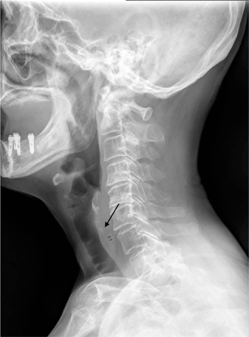
Patient concerns: Case 1, a 75-year-old woman with odynophagia and neck pain occurring 30 min after eating chicken porridge; Case 2, a 32-year-old woman with neck discomfort occurring 2 h after taking a pill; Case 3, a 29-year-old woman reporting FB sensation in the neck that occurred 1 h after eating sausage and rice soup.
Diagnosis: Case 1. Cervical esophageal FB (chicken bone), Case 2. Cervical esophageal FB (oral pill), Case 3. Cervical esophageal FB (food).
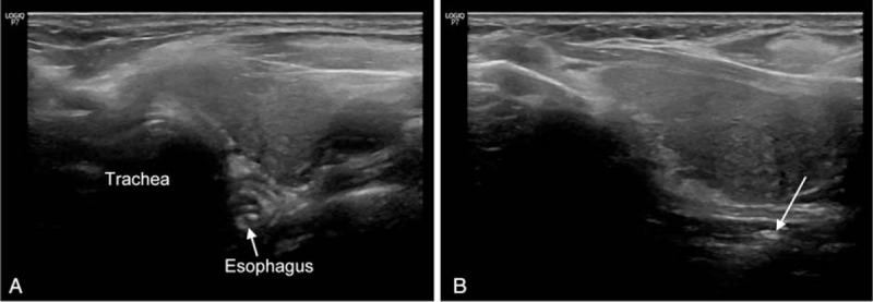
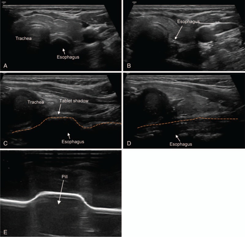
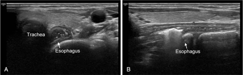
Interventions: Case 1. POCUS, urgent esophagogastroduodenoscopy (EGD) with alligator forceps. POCUS findings; hyperechoic material (suspected FB) that did not disappear by swallowing and esophageal dilatation with pooling of secretions. Case 2. POCUS. POCUS findings; hypoechoic material (suspected FB) that did not disappear by swallowing, and esophageal bulging above the FB, especially observed in the longitudinal view. Case 3. POCUS. POCUS findings; hyperechoic material (suspected FB) with reverberation artifact that did not disappear with swallowing efforts. Prior FB esophageal bulging with persistent air-fluid level was especially observed in the longitudinal view.
Outcomes: Case 1. FB was removed by EGD with alligator forceps. Case 2. Symptoms disappeared under observation without EGD. Follow-up POCUS revealed normalized bulging esophagus. Case 3. These symptoms improved after vomiting a large piece of food material. Three patients were discharged without complications.
Lessons: In this case series, the impacted materials were chicken bone, pill, and food. However, POCUS findings were similar (esophageal dilation, hyperechoic or hypoechoic lesion with mixed echogenic contents in food or secretion, and no change with swallowing efforts). A longitudinal view was useful to assume the presence of cervical esophageal FB in all three cases. Thus, POCUS findings could be indirect signs of a FB in the esophagus.
Conflict of interest statement
The authors have no conflicts of interest to disclose.
Figures
Similar articles
-
Point-of-Care Ultrasound for an Esophageal Foreign Body.J Emerg Med. 2022 Aug;63(2):e53-e56. doi: 10.1016/j.jemermed.2022.04.035. Epub 2022 Jul 21. J Emerg Med. 2022. PMID: 35871027
-
Pediatrician performed point-of-care ultrasound for the detection of ingested foreign bodies: case series and review of the literature.J Ultrasound. 2021 Mar;24(1):107-114. doi: 10.1007/s40477-020-00452-z. Epub 2020 Mar 25. J Ultrasound. 2021. PMID: 32212088 Free PMC article.
-
Bedside sonography for the diagnosis of esophageal food impaction.Am J Emerg Med. 2017 May;35(5):720-724. doi: 10.1016/j.ajem.2017.01.008. Epub 2017 Jan 11. Am J Emerg Med. 2017. PMID: 28119013
-
Point-of-Care Ultrasound for the Diagnosis of Pediatric Foreign Body Ingestion: A Narrative Review and Illustrative Case Report.Pediatr Emerg Care. 2023 Sep 1;39(9):728-733. doi: 10.1097/PEC.0000000000002997. Epub 2023 Jun 21. Pediatr Emerg Care. 2023. PMID: 37339160 Review.
-
Esophageal foreign bodies in adults: systematic review of the literature.Scand J Gastroenterol. 2018 Oct-Nov;53(10-11):1171-1178. doi: 10.1080/00365521.2018.1526317. Epub 2018 Nov 5. Scand J Gastroenterol. 2018. PMID: 30394140
References
-
- Singleton J, Schafer JM, Hinson JS, et al. Bedside sonography for the diagnosis of esophageal food impaction. Am J Emerg Med 2017;35:720–4. - PubMed
-
- Horton LK, Jacobson JA, Powell A, et al. Sonography and radiography of soft-tissue foreign bodies. AJR Am J Roentgenol 2001;176:1155–9. - PubMed
-
- Mori T, Nomura O, Hagiwara Y. Another useful application of point-of-care ultrasound: detection of esophageal foreign bodies in pediatric patients. Pediatr Emerg Care 2019;35:154–6. - PubMed
-
- Jeckovic M, Anupindi SA, Barbir SB, et al. Is ultrasound useful in detection and follow-up of gastric foreign bodies in children? Clin Imaging 2013;37:1043–7. - PubMed
Publication types
MeSH terms
LinkOut - more resources
Full Text Sources
Medical
