. 2020 Jan 24;15(1):e0227634.
doi: 10.1371/journal.pone.0227634. eCollection 2020.

Restoration of Mal overcomes the defects of apoptosis in lung cancer cells

Affiliations

Restoration of Mal overcomes the defects of apoptosis in lung cancer cells

Li-Tao Yang et al. PLoS One. .

Retraction in

Abstract

Background and aims: Cancer is one of the life-threatening diseases of human beings; the pathogenesis of cancer remains to be further investigated. Toll like receptor (TLR) activities are involved in the apoptosis regulation. This study aims to elucidate the role of Mal (MyD88-adapter-like) molecule in the apoptosis regulation of lung cancer (LC) cells.

Methods: The LC tissues were collected from LC patients. LC cells and normal control (NC) cells were isolated from the tissues and analyzed by pertinent biochemical and immunological approaches.

Results: We found that fewer apoptotic LC cells were induced by cisplatin in the culture as compared to NC cells. The expression of Fas ligand (FasL) was lower in LC cells than that in NC cells. FasL mRNA levels declined spontaneously in LC cells. A complex of FasL/TDP-43 was detected in LC cells. LC cells expressed less Mal than NC cells. Activation of Mal by lipopolysaccharide (LPS) increased TDP-43 expression in LC cells. TDP-43 formed a complex with FasL mRNA to prevent FasL mRNA from decay. Reconstitution of Mal or TDP-43 restored the sensitiveness of LC cells to apoptotic inducers.

Conclusions: LC cells express low Mal levels that contributes to FasL mRNA decay through impairing TDP-43 expression. Reconstitution of Mal restores sensitiveness of LC cells to apoptosis inducers that may be a novel therapeutic approach for LC treatment.


Conflict of interest statement

The authors have declared that no competing interests exist.

Figures

Fig 1
Fig 1. Apoptotic defects in LC cells.
The lung cancer (LC) tissues were collected from LC patients (n = 18). LC cells and NC cells (marginal normal tissues; proved by a pathologist) were isolated from the tissues and exposed to cisplatin or saline (vehicle, used as a control) in the culture for 48 h to induce apoptosis. The cells were analyzed by flow cytometry, RT-qPCR and Western blotting. A, gated cells are apoptotic cells. B, summarized data of apoptotic cells in panel A. C, FasL mRNA levels. D, FasL protein levels. E-F, positive correlation between FasL mRNA and apoptotic cells after exposing to cisplatin. Data of bars are presented as mean ± SEM. Each dot in bars presents data obtained from one sample. Statistics: t test.
Fig 2
Fig 2. FasL mRNA decays spontaneously.
NC cells and LC cells were exposed to cisplatin in the culture for 48 h to increase the expression of FasL, washed with fresh medium, and then cultured in the presence or absence of LPS (100 ng/ml). The cells were harvested at indicated timepoints and analyzed by RT-qPCR. The curves show the levels of FasL mRNA in LC cells (A) and NC cells (B). C, results of TLR4 RNAi. TLR4d: TLR4-deficient NC cells. #, TLRd NC cells exposed to LPS in the culture. Data are presented as mean ± SEM and represent 6 independent experiments.
Fig 3
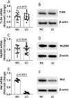
Fig 3. LC cells express less Mal.
LC tissue was collected from LC patients (n = 18). LC cells and NC cells were isolated and analyzed by RT-qPCR and Western blotting. A-B, levels of TLR4. C-D, levels of MyD88. E-F, levels of Mal. Data of bars are presented as mean ± SEM. Each dot in bars presents data obtained from one sample. Statistics: t test.
Fig 4
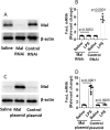
Fig 4. Mal deficiency is associated with FasL mRNA decay.
A-B, NC cells were prepared and exposed to cisplatin in the culture for 48 h. The cells were then washed with fresh medium and treated with Mal RNAi to knock down Mal expression (A). The cells were cultured in the presence or absence of LPS (100 ng/ml) for 48 h. The bars indicate the levels of FasL mRNA (B). C-D, LC cells were prepared; the cells were transfected with Mal-expressing plasmids or control plasmids to restore the expression of Mal (C). The cells were exposed to cisplatin in the culture for 48 h to up regulate the expression of FasL. After washing with fresh medium, the cells were cultured in the presence or absence of LPS (100 ng/ml) for 48 h. The bars indicate the levels of FasL mRNA in the cells. Data of bars are presented as mean ± SEM. Each dot in bars presents data obtained from one sample. Statistics: t test.
Fig 5
Fig 5. Activation of Mal modulates TDP-43 expression.
A-B, lung cancer (LC) tissue was collected from LC patients (n = 18). LC cells and NC cells were isolated from the tissue and analyzed by RT-qPCR and Western blotting. Bars indicate the levels of TDP-43 mRNA (A) and immunoblots indicate TDP-43 protein (B). C-D, scatter dot blots indicate a positive correlation between TDP-43 and Mal in NC and LC cells. E-F, NC cells were cultured in the presence of LPS at indicated concentrations (denoted on the x axis of E) for 48 h. Bars indicate the mRNA levels of TDP-43. Immunoblots indicate the protein levels of TDP-43. G-H, HC cells and LC cells were prepared and analyzed by RNA-immunoprecipitation assay; a complex of FasL mRNA and TDP-43 protein was identified. Bars indicate FasL mRNA in the complex. Immunoblots indicate TDP-43 protein in the complex. I, results of TDP-43 RNAi. J, NC cells (with or without TDP-43 depletion) were exposed to cisplatin in the culture for 48 h. Bars show FasL mRNA expression in EC cells. K, results of TDP-43 restoration by transfection of TDP-43 expressing plasmids (TDP-43R). L, Bars show FasL mRNA expression in LC cells. Data of bars are presented as mean ± SEM. Each dot in bars presents data obtained from one sample. Statistics: A and G, t test. E, ANOVA: p<0.0001; *p<0.01, compared with the group “0” (Bonferroni test). J and L, ANOVA + Dunnett’s test. Data of B represent 6 independent experiments (protein extracts of 18 samples were pooled). Data E-H represent 6 independent experiments. #, cells were treated with Mal RNAi to knock down the expression of Mal. $, cells were treated with control RNAi.
Fig 6
Fig 6. Restoration of apoptotic machinery in LC cells by modulation of Mal or TDP-43 expression.
A-B, NC cells were prepared and treated with Mal RNAi or TDP-43 RNAi to knock down the expression of Mal or TDP-43. The cells were exposed to cisplatin in the culture for 48 h. The gated dot plots show apoptotic cells. The bars show summarized data of apoptotic cells. C-D, LC cells were prepared and transfected with Mal-expressing or TDP-43-expressing plasmids as denoted above each subpanel. The cells were exposed to cisplatin in the culture for 48 h. The gated dot plots show apoptotic cells. The bars show summarized data of apoptotic cells. Data of bars are presented as mean ± SEM. Each dot in bars presents data obtained from one sample. The data represent 6 independent experiments. Statistics: ANOVA + Bonferroni test.

References

    1. Nanavaty P, Alvarez MS, Alberts WM. Lung cancer screening: advantages, controversies, and applications. Cancer Control. 2014;21(1):9–14. Epub 2013/12/21. 10.1177/107327481402100102 . - DOI - PubMed
    1. Kadara H, Scheet P, Wistuba II, Spira AE. Early Events in the Molecular Pathogenesis of Lung Cancer. Cancer Prev Res (Phila). 2016;9(7):518–27. Epub 2016/03/24. 10.1158/1940-6207.Capr-15-0400 . - DOI - PubMed
    1. Hirsch FR, Scagliotti GV, Mulshine JL, Kwon R, Curran WJ Jr., Wu YL, et al. Lung cancer: current therapies and new targeted treatments. Lancet. 2017;389(10066):299–311. Epub 2016/08/31. 10.1016/S0140-6736(16)30958-8 . - DOI - PubMed
    1. Zhang L, Yu J. Role of apoptosis in colon cancer biology, therapy, and prevention. Curr Colorectal Cancer Rep. 2013;9(4). Epub 2013/11/26. 10.1007/s11888-013-0188-z - DOI - PMC - PubMed
    1. Pore MM, Hiltermann TJ, Kruyt FA. Targeting apoptosis pathways in lung cancer. Cancer Lett. 2013;332(2):359–68. Epub 2010/10/27. 10.1016/j.canlet.2010.09.012 . - DOI - PubMed

Publication types

MeSH terms