

An IL-18-centered inflammatory network as a biomarker for cerebral white matter injury
- PMID: 31978079
- PMCID: PMC6980497
- DOI: 10.1371/journal.pone.0227835
An IL-18-centered inflammatory network as a biomarker for cerebral white matter injury
Abstract
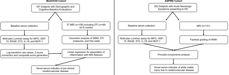
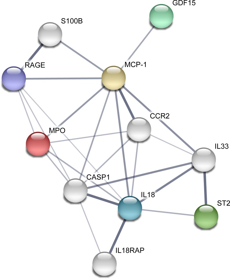
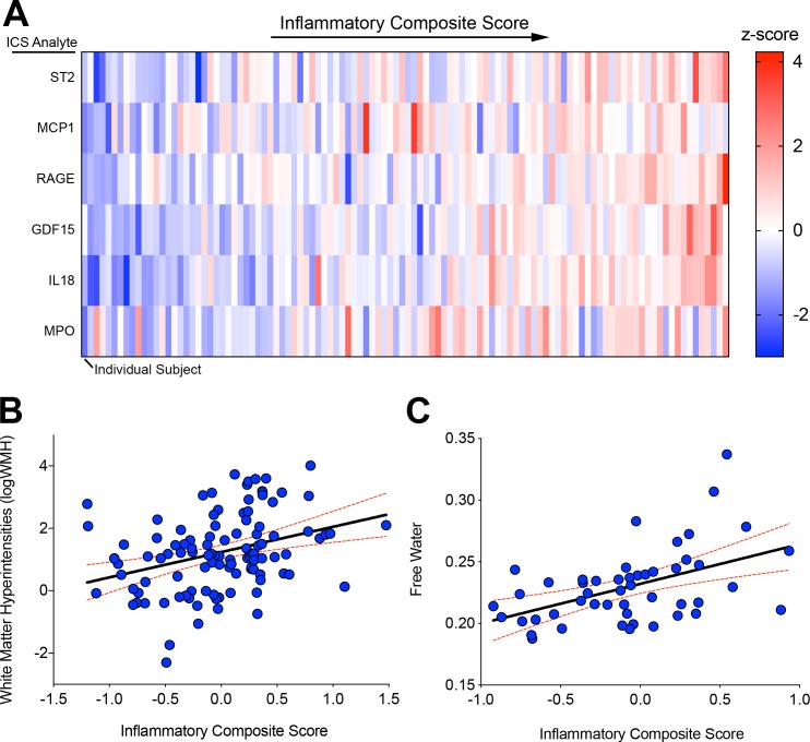
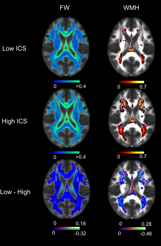
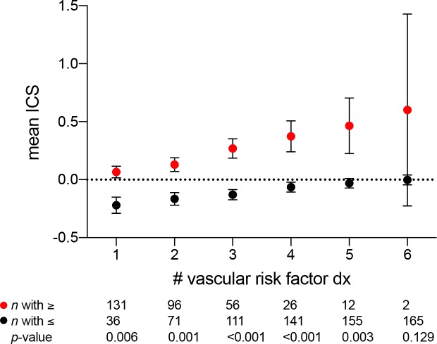
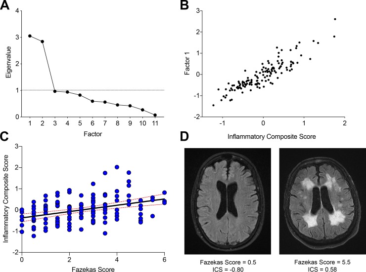
Chronic systemic sterile inflammation is implicated in the pathogenesis of cerebrovascular disease and white matter injury. Non-invasive blood markers for risk stratification and dissection of inflammatory molecular substrates in vivo are lacking. We sought to identify whether an interconnected network of inflammatory biomarkers centered on IL-18 and all previously associated with white matter lesions could detect overt and antecedent white matter changes in two populations at risk for cerebral small vessel disease. In a cohort of 167 older adults (mean age: 76, SD 7.1, 83 females) that completed a cognitive battery, physical examination, and blood draw in parallel with MR imaging including DTI, we measured cerebral white matter hyperintensities (WMH) and free water (FW). Concurrently, serum levels of a biologic network of inflammation molecules including MPO, GDF-15, RAGE, ST2, IL-18, and MCP-1 were measured. The ability of a log-transformed population mean-adjusted inflammatory composite score (ICS) to associate with MR variables was demonstrated in an age and total intracranial volume adjusted model. In this cohort, ICS was significantly associated with WMH (β = 0.222, p = 0.013), FW (β = 0.3, p = 0.01), and with the number of vascular risk factor diagnoses (r = 0.36, p<0.001). In a second cohort of 131 subjects presenting for the evaluation of acute neurologic deficits concerning for stroke, we used serum levels of 11 inflammatory biomarkers in an unbiased principal component analysis which identified a single factor significantly associated with WMH. This single factor was strongly correlated with the six component ICS identified in the first cohort and was associated with WMH in a generalized linear regression model adjusted for age and gender (p = 0.027) but not acute stroke. A network of inflammatory molecules driven by IL-18 is associated with overt and antecedent white matter injury resulting from cerebrovascular disease and may be a promising peripheral biomarker for vascular white matter injury.
Conflict of interest statement
Dr. DeCarli serves as a consultant to Novartis Pharmaceuticals on a trial studying the safety of heart failure medication. The University of California has filed U.S. patent (16/487,332) application for: “Serologic assay for silent brain ischemia” for which Drs. Hinman and Xiao are co-inventors. This does not alter our adherence to PLOS ONE policies on sharing data and materials.
Figures
References
-
- Uiterwijk R, van Oostenbrugge RJ, Huijts M, De Leeuw PW, Kroon AA, Staals J. Total Cerebral Small Vessel Disease MRI Score Is Associated with Cognitive Decline in Executive Function in Patients with Hypertension. Front Aging Neurosci. 2016;8:301 Epub 2016/12/27. 10.3389/fnagi.2016.00301 - DOI - PMC - PubMed
-
- Staszewski J, Piusinska-Macoch R, Skrobowska E, Brodacki B, Pawlik R, Dutkiewicz T, et al. Significance of Haemodynamic and Haemostatic Factors in the Course of Different Manifestations of Cerebral Small Vessel Disease: The SHEF-CSVD Study-Study Rationale and Protocol. Neurosci J. 2013;2013:424695 Epub 2013/01/01. 10.1155/2013/424695 - DOI - PMC - PubMed
Publication types
MeSH terms
Substances
Grants and funding
- R01 NS017950/NS/NINDS NIH HHS/United States
- R01 AG054076/AG/NIA NIH HHS/United States
- R01 AG048234/AG/NIA NIH HHS/United States
- P01 AG019724/AG/NIA NIH HHS/United States
- P30 AG010129/AG/NIA NIH HHS/United States
- UH3 NS100608/NS/NINDS NIH HHS/United States
- T35 AG026736/AG/NIA NIH HHS/United States
- UH2 NS100608/NS/NINDS NIH HHS/United States
- RF1 NS130659/NS/NINDS NIH HHS/United States
- RF1 AG032289/AG/NIA NIH HHS/United States
- T32 AG023481/AG/NIA NIH HHS/United States
- P30 AG062422/AG/NIA NIH HHS/United States
- P30 AG066546/AG/NIA NIH HHS/United States
- R01 AG032289/AG/NIA NIH HHS/United States
- U01 AG052409/AG/NIA NIH HHS/United States
LinkOut - more resources
Full Text Sources
Other Literature Sources
Medical
Research Materials
Miscellaneous
