. 2020 Jan 24;15(1):e0228011.
doi: 10.1371/journal.pone.0228011. eCollection 2020.

Metabolic and lipidomic profiling of steatotic human livers during ex situ normothermic machine perfusion guides resuscitation strategies

Affiliations

Metabolic and lipidomic profiling of steatotic human livers during ex situ normothermic machine perfusion guides resuscitation strategies

Siavash Raigani et al. PLoS One. .

Abstract

There continues to be a significant shortage of donor livers for transplantation. One impediment is the discard rate of fatty, or steatotic, livers because of their poor post-transplant function. Steatotic livers are prone to significant ischemia-reperfusion injury (IRI) and data regarding how best to improve the quality of steatotic livers is lacking. Herein, we use normothermic (37°C) machine perfusion in combination with metabolic and lipidomic profiling to elucidate deficiencies in metabolic pathways in steatotic livers, and to inform strategies for improving their function. During perfusion, energy cofactors increased in steatotic livers to a similar extent as non-steatotic livers, but there were significant deficits in anti-oxidant capacity, efficient energy utilization, and lipid metabolism. Steatotic livers appeared to oxidize fatty acids at a higher rate but favored ketone body production rather than energy regeneration via the tricyclic acid cycle. As a result, lactate clearance was slower and transaminase levels were higher in steatotic livers. Lipidomic profiling revealed ω-3 polyunsaturated fatty acids increased in non-steatotic livers to a greater extent than in steatotic livers. The novel use of metabolic and lipidomic profiling during ex situ normothermic machine perfusion has the potential to guide the resuscitation and rehabilitation of steatotic livers for transplantation.


Conflict of interest statement

Dr. Uygun is inventor on pending patents relevant to this study (WO/2011/002926; WO/2011/35223) and has a provisional patent application relevant to this study (MGH 22743). Dr. Uygun has a financial interest in Organ Solutions, a company focused on developing organ preservation technology. Dr. Uygun’s interests are managed by the MGH and Partners HealthCare in accordance with their conflict of interest policies. The hemoglobin-based oxygen carrier, Hemopure was kindly provided by HbO2 Therapeutics. This does not alter our adherence to PLOS ONE policies on sharing data and materials.

Figures

Fig 1
Fig 1. Vascular resistance during perfusion of steatotic and non-steatotic livers.
Steatotic livers tend to have higher vascular resistance at initiation of perfusion, which improves with time. No significant differences were observed between the groups with mixed model analysis. (a) Hepatic artery resistance and (b) Portal vein resistance are shown during 3 hours of normothermic machine perfusion. Data shown as mean ± SEM. ST, steatotic; NST, non-steatotic.
Fig 2
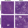
Fig 2. Histologic differences between steatotic and non-steatotic livers before and after perfusion.
Representative H&E-stained liver sections from steatotic and non-steatotic livers shown pre-perfusion and post-perfusion (3 hours of normothermic machine perfusion). Steatotic liver biopsies demonstrate large lipid macrodroplets in the cytoplasm of hepatocytes. No significant change in macrosteatosis content was observed after three hours of perfusion in steatotic and non-steatotic livers.
Fig 3
Fig 3. Characterization of liver function and injury during perfusion.
(a) Release of alanine aminotransferase. ALT was higher in steatotic livers but did not reach significance. (b) Bile production measured at each hour of perfusion. Bile production increased at comparable volumes between groups. (c) Lactate content measured in the venous outflow of livers during perfusion. ST livers have higher lactate content and slower clearance compared to NST livers. (d) Glucose content measured in the venous outflow. ST livers release higher levels of glucose during perfusion compared to NST livers. * indicates P<0.05 for random intercept mixed model comparison of repeated measures data between groups. Comparisons are made at each time point between groups with respect to the measured change from time = 0 minutes. Data shown as mean ± SEM. ST, steatotic; NST, non-steatotic.
Fig 4
Fig 4. Redox factor changes in steatotic and non-steatotic livers during perfusion.
Ratio of metabolite concentration fold change at each hour of perfusion compared to pre-perfusion concentration shown for (a) reduced glutathione, (b) oxidized glutathione, and (c) N-acetylcysteine. (d) Concentration of nitric oxide in perfusate samples. Steatotic livers demonstrate significant deficits in redox capacity and NO synthesis during perfusion. ** indicates P≤0.05, * indicates 0.05
Fig 5
Fig 5. Analysis of energy cofactor changes in steatotic and non-steatotic liver tissue during perfusion.
(a) Measured ATP:ADP ratio, (b) ATP:AMP ratio, (c) energy charge, and (d) NADPH:NADP+ ratios at each hour of perfusion. Energy charge was calculated as (2*ATP + ADP)/(ATP + ADP + AMP). Within group comparisons at 60, 120, and 180 minutes are made to a pre-perfusion measurement (time = 0 min). * indicates P<0.05 for random intercept mixed model comparison of repeat measures data to pre-perfusion levels. Data shown are mean ± SEM. ST, steatotic; NST, non-steatotic.
Fig 6
Fig 6. Metabolic capacity of fatty acid oxidation in steatotic and non-steatotic livers during perfusion.
(a) Carnitine and (b) β-hydroxybutyrate concentration fold change at each hour of perfusion compared to pre-perfusion concentration. (c) Heatmap of fatty acylcarnitine metabolite concentration fold change at each hour of perfusion. Steatotic livers demonstrate upregulation of fatty acid oxidation, with resultant depletion of carnitine and fatty acylcarnitine levels during perfusion, but favor greater ketone synthesis compared to non-steatotic livers. ** indicates P≤0.05, * indicates 0.05
Fig 7
Fig 7. Metabolic analysis of cellular metabolism in steatotic and non-steatotic livers during perfusion.
Ratio of metabolite concentration fold change at each hour of perfusion compared to pre-perfusion concentration shown for (a) tricyclic acid cycle, (b) glycolysis, and (c) pentose phosphate pathway. Steatotic livers demonstrate decreased flux through each metabolic pathway. ** indicates P≤0.05, * indicates 0.05
Fig 8
Fig 8. ω-3 fatty acid lipidomic profiles of steatotic and non-steatotic livers during perfusion.
Ratio of metabolite concentration fold change at each hour of perfusion compared to pre-perfusion concentration shown for the free fatty acid (FFA) form of (a) eicosapentaenoic acid (EPA), (b) docosapentaenoic acid (DPA), and (c) docosahexaenoic acid (DHA). (d) Heatmap of EPA, DPA, and DHA concentration fold changes in cholesterol ester (CE), FFA, monoacylglycerol (MAG), diacylglycerol (DAG), and triacylglycerol (TAG) forms. ** indicates P≤0.05, * indicates 0.05
Fig 9
Fig 9. Bile acid profiles of steatotic and non-steatotic livers during perfusion.
Ratio of metabolite concentration fold change at each hour of perfusion compared to pre-perfusion concentration shown for (a) cholesterol, (b) taurine, and (c) choline. (d) Heatmap of primary and secondary bile acid concentration fold changes during perfusion. ** indicates P≤0.05, * indicates 0.05

References

    1. United Network for Organ Sharing (UNOS). Available from: https://unos.org/data/transplant-trends/.
    1. Moosburner S, Gassner J, Nosser M, Pohl J, Wyrwal D, Claussen F, et al. Prevalence of Steatosis Hepatis in the Eurotransplant Region: Impact on Graft Acceptance Rates. HPB Surg. 2018;2018:6094936 Epub 2018/12/06. 10.1155/2018/6094936 - DOI - PMC - PubMed
    1. Chu MJ, Dare AJ, Phillips AR, Bartlett AS. Donor Hepatic Steatosis and Outcome After Liver Transplantation: a Systematic Review. J Gastrointest Surg. 2015;19(9):1713–24. Epub 2015/04/29. 10.1007/s11605-015-2832-1 . - DOI - PubMed
    1. Selzner M, Clavien PA. Fatty liver in liver transplantation and surgery. Semin Liver Dis. 2001;21(1):105–13. Epub 2001/04/12. 10.1055/s-2001-12933 . - DOI - PubMed
    1. Farrell GC, Teoh NC, McCuskey RS. Hepatic microcirculation in fatty liver disease. Anat Rec (Hoboken). 2008;291(6):684–92. Epub 2008/05/20. 10.1002/ar.20715 . - DOI - PubMed

Publication types