Review
. 2020 Apr:137:104760.
doi: 10.1016/j.nbd.2020.104760. Epub 2020 Jan 21.

Immunotherapy for Parkinson's disease

Affiliations
Review

Immunotherapy for Parkinson's disease

Aaron D Schwab et al. Neurobiol Dis. 2020 Apr.

Abstract

With the increasing prevalence of Parkinson's disease (PD), there is an immediate need to interdict disease signs and symptoms. In recent years this need was met through therapeutic approaches focused on regenerative stem cell replacement and alpha-synuclein clearance. However, neither have shown long-term clinical benefit. A novel therapeutic approach designed to affect disease is focused on transforming the brain's immune microenvironment. As disordered innate and adaptive immune functions are primary components of neurodegenerative disease pathogenesis, this has emerged as a clear opportunity for therapeutic development. Interventions that immunologically restore the brain's homeostatic environment can lead to neuroprotective outcomes. These have recently been demonstrated in both laboratory and early clinical investigations. To these ends, efforts to increase the numbers and function of regulatory T cells over dominant effector cells that exacerbate systemic inflammation and neurodegeneration have emerged as a primary research focus. These therapeutics show broad promise in affecting disease outcomes beyond PD, such as for Alzheimer's disease, stroke and traumatic brain injuries, which share common neurodegenerative disease processes.

Keywords: Alzheimer’s disease; Effector T cells; Granulocyte-macrophage colony stimulating factor; Immune homeostasis; Immune transformation; Ischemic stroke; Neurodegeneration; Neurodegenerative disorders; Neuroinflammation; Neuroprotection; Nigrostriatal degeneration; Parkinson’s disease; Regulatory T cells; Teffs; Traumatic brain injury; Tregs.


Conflict of interest statement

Declaration of Competing Interest None

Figures

Fig. 1.
Fig. 1.
Immunity and neurodegenerative disease pathogenesis. Neurodegenerative processes often involve interactions between infiltrating effector T cells (Teffs) and microglia to affect the progression of neurodegenerative disease either due to aberrant protein processing or danger/damage signaling due to neuronal injury and death. Microglia respond to a reactive phenotype during cell-cell interactions with Teffs after migrating across the blood brain barrier (BBB). While the pathological consequences and origins of such neuroinflammation varies across disease states, the inflammatory signature shared by the represented neurodegenerative diseases identifies a common target for disease therapy. (A) In Parkinson’s disease, α-syn is modified, misfolded, oligomerized, and released into the extracellular environment following neuronal injury. Misfolded and oxidatively modified α-syn aggregates and elicits microglial activation that perpetuates neuronal damage within the substantia nigra pars compacta (SNc). Modified self-proteins, such as aggregated N-α-syn with proinflammatory milieu, initiate a systemic, adaptive immune response following their drainage to secondary lymphoid tissues. Increased BBB permeability and Teff influx exacerbate neuroinflammation and neurodegeneration upon recognition in the CNS through the secretion of inflammatory mediators that shift the brain microenvironment towards a pro-inflammatory phenotype. (B) In Alzheimer’s disease, inflammation-associated neuronal death results in the release of amyloid-β (Aβ) plaques and neurofibrillary tangles (NFTs) into the extracellular environment which induce inflammation and subsequent neural death. Increased inflammation augments aberrant amyloid protein processing, Aβ accumulation, and hyperphosphorylation of Tau. (C) In TBI, trauma to the brain causes tissue damage, which in turn activates microglia and induces Teff influx. Microglia shift from a homeostatic to a reactive phenotype with secretion of proinflammatory factors leading to increased overall cytotoxicity that propagates neuroinflammation and neurodegeneration. (D) Following an ischemic stroke, tissue damage and BBB injury initiate microglial activation and influx of pro-inflammatory immune cells. Teffs infiltrating the brain, secrete neurotoxic and inflammatory mediators alongside reactive microglia resulting in penumbric spreading of neuronal death and astroglial scarring.
Fig. 2.
Fig. 2.
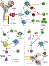
Therapeutic transformation of the brain’s microenvironment in neurodegenerative disease. A neuroinflammatory microenvironment heralds the onset and progression of neurodegenerative disease. Inflammation perpetuates microglial activation and subsequent neurodegeneration and is pivotal in the progression of Parkinson’s disease, Alzheimer’s disease, stroke, and traumatic brain injury. In most neurodegenerative disorders, aberrant protein modifications and misfolding allow fibrillogenic and aggregated forms to be released into the extracellular environment. Similarly in TBI and stroke, damage/danger signals from tissues and neurons are also produced and released upon injury. Modified proteins and DAMPs initiate the activation of microglia with the production and release of inflammatory mediators and cytokines as well as reactive oxygen and nitrogen species. These prove to be neurotoxic with the ability to damage surrounding neurons. Additionally, the proinflammatory milieu drains to secondary lymphoid tissues where it activates antigen presenting cells (APCs) to present modified self-antigens such as nitrated α-synuclein. Under the influence of proinflammatory co-stimulatory signals, naïve T cells initiate programs to differentiate and expand into pro-inflammatory effector T cells (Teffs) such as Th1 and Th17 cells. These Teffs extravasate across the blood brain barrier at inflammatory foci whereby Teffs are reactivated by microglia or macrophages which exacerbate neuroinflammation and neurodegeneration through the secretion of inflammatory mediators. Overall, Teff-microglia interactions shift the brain microenvironment towards a pro-inflammatory neurotoxic environment that hastens disease progression. On the other hand, regulatory T cells (Tregs) have the capacity to harness microglial and APC activation, attenuate inflammation, inhibit Teff induction, and induce astrocytic neurotrophins, thus effectively transforming a neurotoxic environment to a neurotrophic state. In chronic neurodegenerative disorders and acute CNS damage, Treg processes are often overwhelmed due to their low numbers or dysfunction. Therefore immune modulating agents, such as anti-CD3 mAbs, vasoactive intestinal peptide (VIP), or granulocyte macrophage colony stimulating factor (GM-CSF) are utilized to increase Treg number and function which also can extravasate at sites of neuroinflammation and execute Treg processes to rebalance the neurotoxic state to one of neuroprotection.
Fig. 3.
Fig. 3.
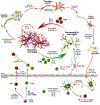
Mechanisms underlying Treg expansion. The growing body of research that implicates immunological dysfunction in the pathology of Parkinson’s disease and other neurodegenerative disorders has shed light on new pharmacological targets capable of addressing more causal aspects of the neurodegenerative disease. Specifically, Treg inducing agents capable of shifting the immune environment towards an anti-inflammatory, neuroprotective state are being explored for future clinical application. (A) GM-CSF acts as a Treg inducing agent by initially facilitating the differentiation of bone marrow progenitor cells into tolerogenic dendritic cells (DCs). These dendritic cells express the surface markers OX40L and Jagged-1 (Jag-1) which induce and expand natural Treg and induce Treg proliferation following interactions with their cognate receptors. TGF-β secreted by the BMDCs enable the conversion of Teffs into Tregs in the presence of co-stimulatory molecules. A specific population of tolerogenic DCs that are CD8α- are remarkably responsive to GM-CSF as they expand in both number and function and directly transform Teffs to Tregs. (B) In contrast to other immunomodulating therapies, anti-CD3 monoclonal antibodies (mAbs) directly affect peripheral T cells. Upon anti-CD3 mAb binding the CD3/TCR complex of a naïve T cell, it is capable of shedding or internalizing the receptor/antibody complex or propagating a signaling cascade. This signaling can result in either induction of an anergic program capable of transient immune suppression, or an apoptotic program by the activated cell. Apoptosis causes the release of apoptotic bodies which secrete TGF-β into the environment or are phagocytosed by macrophages which then releases TGF-β into the extracellular space. TGF-β shifts DCs into a tolerogenic state or acts on naïve T cells to induce Tregs. (C) VIP generates tolerogenic DCs from myeloid specific bone marrow progenitors. VIP-differentiated tolerogenic DCs induce T regulatory 1 (Tr1) cells from CD4+ CD25− naïve T cells which secrete anti-inflammatory cytokines such as TGF-β and IL-10. Within an environment associated with factors such as IL-10, TGF-β, and INF-α, VIP facilitates the differentiation of naïve T cells into Tregs with high levels of CTLA-4 on the cell surface. In the presence of VIP, Th3 cells are also induced from the CD4+ CD25− compartment, furthering the anti-inflammatory shift in the microenvironment via the secretion of TGF-β.

References

    1. Allen Reish HE, Standaert DG, 2015. Role of alpha-synuclein in inducing innate and adaptive immunity in Parkinson disease. J Parkinsons Dis. 5, 1–19. - PMC - PubMed
    1. Anthonymuthu TS, Kenny EM, Bayir H, 2016. Therapies targeting lipid peroxidation in traumatic brain injury. Brain Res. 1640, 57–76. - PMC - PubMed
    1. Bach JF, 2011. Anti-CD3 antibodies for type 1 diabetes: beyond expectations. Lancet. 378, 459–460. - PubMed
    1. Bachiller S, Jimenez-Ferrer I, Paulus A, Yang Y, Swanberg M, Deierborg T, Boza-Serrano A, 2018. Microglia in neurological diseases: a road map to brain-disease dependent-inflammatory response. Front Cell Neurosci. 12, 488. - PMC - PubMed
    1. Banerjee K, Kumar S, Ross KA, Gautam S, Poelaert B, Nasser MW, Aithal A, Bhatia R, Wannemuehler MJ, Narasimhan B, Solheim JC, Batra SK, Jain M, 2018. Emerging trends in the immunotherapy of pancreatic cancer. Cancer Lett. 417, 35–46. - PMC - PubMed

Publication types