Randomized Controlled Trial
. 2020:25:102182.
doi: 10.1016/j.nicl.2020.102182. Epub 2020 Jan 14.

Hippocampal plasticity underpins long-term cognitive gains from resistance exercise in MCI

Affiliations
Randomized Controlled Trial

Hippocampal plasticity underpins long-term cognitive gains from resistance exercise in MCI

Kathryn M Broadhouse et al. Neuroimage Clin. 2020.

Abstract

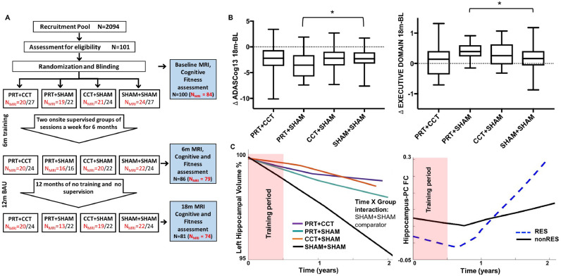
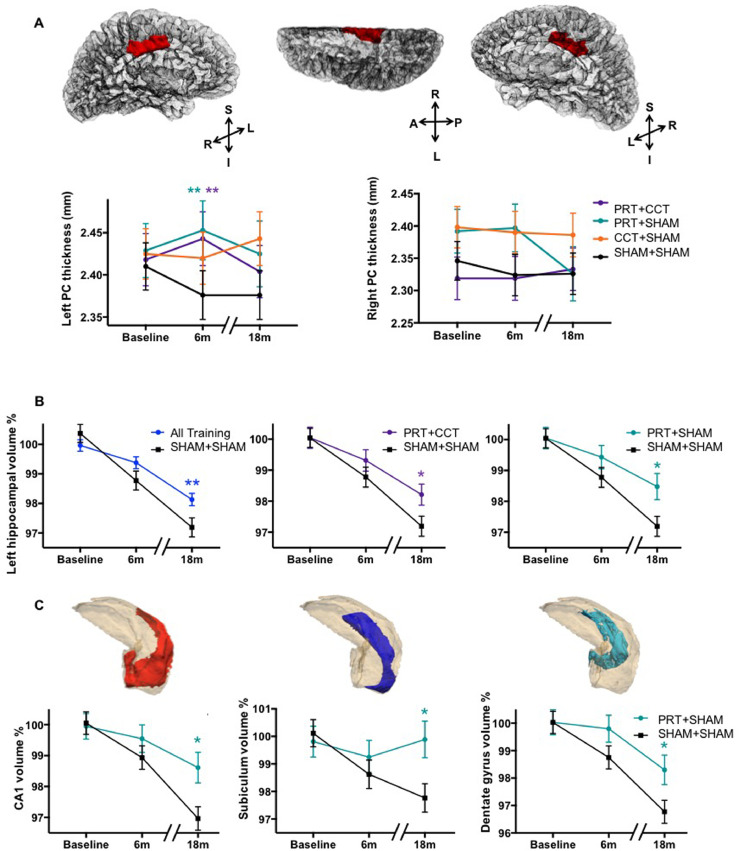
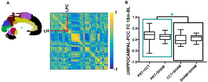
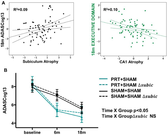
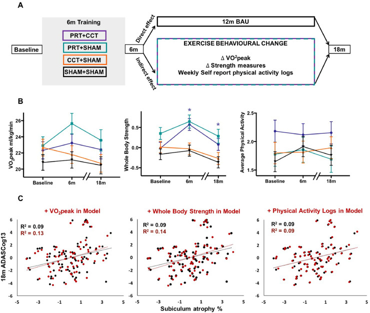
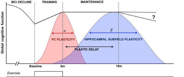
Dementia affects 47 million individuals worldwide, and assuming the status quo is projected to rise to 150 million by 2050. Prevention of age-related cognitive impairment in older persons with lifestyle interventions continues to garner evidence but whether this can combat underlying neurodegeneration is unknown. The Study of Mental Activity and Resistance Training (SMART) trial has previously reported within-training findings; the aim of this study was to investigate the long-term neurostructural and cognitive impact of resistance exercise in Mild Cognitive Impairment (MCI). For the first time we show that hippocampal subareas particularly susceptible to volume loss in Alzheimer's disease (AD) are protected by resistance exercise for up to one year after training. One hundred MCI participants were randomised to one of four training groups: (1) Combined high intensity progressive resistance and computerised cognitive training (PRT+CCT), (2) PRT+Sham CCT, (3) CCT+Sham PRT, (4) Sham physical+sham cognitive training (SHAM+SHAM). Physical, neuropsychological and MRI assessments were carried out at baseline, 6 months (directly after training) and 18 months from baseline (12 months after intervention cessation). Here we report neuro-structural and functional changes over the 18-month trial period and the association with global cognitive and executive function measures. PRT but not CCT or PRT+CCT led to global long-term cognitive improvements above SHAM intervention at 18-month follow-up. Furthermore, hippocampal subfields susceptible to atrophy in AD were protected by PRT revealing an elimination of long-term atrophy in the left subiculum, and attenuation of atrophy in left CA1 and dentate gyrus when compared to SHAM+SHAM (p = 0.023, p = 0.020 and p = 0.027). These neuroprotective effects mediated a significant portion of long-term cognitive benefits. By contrast, within-training posterior cingulate plasticity decayed after training cessation and was unrelated to long term cognitive benefits. Neither general physical activity levels nor fitness change over the 18-month period mediated hippocampal trajectory, demonstrating that enduring hippocampal subfield plasticity is not a simple reflection of post-training changes in fitness or physical activity participation. Notably, resting-state fMRI analysis revealed that both the hippocampus and posterior cingulate participate in a functional network that continued to be upregulated following intervention cessation. Multiple structural mechanisms may contribute to the long-term global cognitive benefit of resistance exercise, developing along different time courses but functionally linked. For the first time we show that 6 months of high intensity resistance exercise is capable of not only promoting better cognition in those with MCI, but also protecting AD-vulnerable hippocampal subfields from degeneration for at least 12 months post-intervention. These findings emphasise the therapeutic potential of resistance exercise; however, future work will need to establish just how long-lived these outcomes are and whether they are sufficient to delay dementia.

Keywords: Hippocampus; Mild cognitive impairment; Plasticity; Randomised controlled trial; Resistance exercise; Subfields.


Conflict of interest statement

Declaration of Competing Interest The authors have no conflicts of interest to declare.

Figures

Fig 1
Fig. 1
SMART Trial design and statistical models. (A) Randomization was into one of four training groups – combined progressive resistance training and computerised cognitive training (PRT+CCT), PRT and cognitive sham (PRT+SHAM), CCT and sham exercise (CCT+SHAM), sham exercise and cognitive sham, (SHAM+SHAM). MRI, cognitive and fitness assessments were carried out at baseline, directly after 6 months of training (6M) and at 18 months (18M), following a one-year usual care (UC) period with weekly telephone contact by research staff to administer health/adverse event checks in all participants. In addition, cognitive, physical, social, recreational, volunteer and religious activity participation was recorded daily for the entire 18 months in a logbook by each participant. (B) Raw change in the trial's primary outcome (ADASCog error scores) over the complete 18-month period, as well as neuropsychologically-defined Executive domain scores were compared between SHAM+SHAM and the three training groups. Box-and-Whisker plots show the median (horizontal line), interquartile range (box) and upper and lower quartiles (whiskers) of change in outcome scores for each training group. Dotted line indicates no change in outcome over the 18-month period. LME analysis found significantly (*Time x Group p < 0.05 superimposed on the Box-and-Whisker plots) improved cognition in the PRT+SHAM group compared to SHAM+SHAM on both outcomes (model including covariates age, sex and education, as well as group factor and time as continuous variable). (C) Basic Linear Mixed Effects (LME) model including baseline covariates (age, sex, education), time (as continuous variable), group (SHAM+SHAM comparator) and Time x Group interaction. Unadjusted locally-weighted mean trajectories were plotted over a sliding temporal window (i.e., lowess plot) for hippocampal volume as percentage of baseline and functional connectivity z-scores for all four groups over the 18-month trial period. Temporal relationship determined the subsequent modelled time interaction function in the LME.
Fig 2
Fig. 2
Resistance exercise preserves and protects Alzheimer-vulnerable hippocampal subfields for up to one year after training. (A) Left but not right PC cortical thickness was enhanced by any resistance exercise training as opposed to double-sham; however, this advantage was lost one year after training cessation. (**) refers to significant within-training effect of PRT+CCT vs SHAM+SHAM (F = 11.1, p = 0.005 FDR corrected, DF = 39.2) and PRT+SHAM vs SHAM+SHAM (F = 15.2, p = 0.005 FDR corrected, DF = 36.4), using an identical LME model restricted to baseline and 6 months. No Time x Group effects were observed when testing the complete 18-month model. (B) Left hippocampal volume trajectory is protected from atrophy by any training compared to double-sham (left; Time x Group F = 7.27, p = 0.008, DF = 157.9), the effect specific to groups that included resistance training (PRT+CCT: middle, F = 5.10, p = 0.041 FDR corrected, DF = 84.0; and PRT+SHAM: F = 7.22, p = 0.027 FDR corrected, DF = 79.6). (C) Resistance exercise alone protects Alzheimer-vulnerable hippocampal subfields, including: left CA1 (F = 8.81, p = 0.020 FDR corrected, DF = 81.0); Subiculum (F = 7.14, p = 0.023 FDR corrected, DF = 80.2); and dentate gyrus (F = 5.53, p = 0.035 FDR corrected, DF = 79.1). For each of these subfields, 6 months of resistance exercise protected participants from 2% to 3% volumetric loss over the complete 18-month follow-up period. Means (±SEM) adjusted for baseline age, years of education, sex, and baseline volume normalized by ICV. (*) refers to significant Time x Group interaction at p < 0.05. NB: Idealised time points (baseline, 6M, 18M) are displayed for visualisation purposes only; statistics are solely from LME models where real-time is treated as a continuous variable.
Fig 3
Fig. 3
Resistance exercise leads to long term increase in functional connectivity between left hippocampus and posterior cingulate. (A) An individual functional connectome from one SMART participant at baseline showing the correlation between all 116 AAL-atlas brain regions (left) in the form of a correlation heat map (right). Individual connectomes were generated at each time point, and correlation values between the left hippocampus and PC regions extracted to investigate the change in functional connectivity using our LME model as outlined in Fig 1E. (B) Raw change in the functional connectivity between left hippocampus and PC over the complete 18-month period is depicted at the individual level. Box-and-Whisker plots show the median (horizontal line), interquartile range (box) and upper and lower quartiles (whiskers) of change in functional connectivity between left PCC and hippocampus for each training group. LME analysis found significantly increased connectivity for those two groups that underwent resistance exercise training (PRT+CCT and PRT+SHAM) compared to those that did not (CCT+SHAM and SHAM+SHAM). *Time x Group p < 0.05; model included covariates age, sex and education, as well as group factor and time as continuous variable.
Fig 4
Fig. 4
Therapeutic benefits of resistance exercise on cognition one year after training are mediated by preservation of AD-vulnerable hippocampal subfields. (A) Multiple regression analysis tested the unique variance accounted in the primary outcome (ADASCog) and Executive function by 18-month subiculum or CA1 atrophy (as percent change from baseline; age, sex and education were also in the model). Partial plots here show that subiculum atrophy was an independent predictor of ADAGCog13, whilst CA1 atrophy was an independent predictor of Executive function. The beta values for each of these (β = 0.28 and −0.25, respectively) were of similar magnitude to clinically meaningful predictors age (β = 0.22 and −0.30) and education (β = −0.24 and 0.53). (B) Since resistance exercise protects and preserves subiculum and CA1 volume (Fig 2C), is therapeutically effective on cognition in the long term (A), and the former predict the latter (B), we re-ran the cognitive LME model in (A) with 18-month change in subiculum volume (Δsubic; expressed as percentage change from baseline) as an additional covariate. This abolished the therapeutic effect of resistance exercise on ADASCog. Accordingly, resistance exercise-dependent plasticity of the subiculum mediates long-term therapeutic benefits on global cognition.
Fig 5
Fig. 5
Physical fitness and physical activity behaviour after training does not mediate long-term protection of hippocampal subfields. (A) A conceptual schema showing how the long-term effects of resistance exercise on the brain could arise directly following training in the context of reverting to business-as-usual (BAU) or could be mediated by changes in physical fitness or physical activity behaviour unrelated to the interventions. We modelled exercise-related behaviour and physical fitness in three different ways, using long term changes in physiologic VO2peak and whole-body muscle strength measurements, as well as daily physical activity logs over 18 months. (B) Change in fitness and exercise behaviour is charted over time (mean (±SEM)) using a LME model, revealing a significant Time x Group (*) effect of PRT+CCT on whole body strength during training and over the complete 18-month period compared to SHAM+SHAM (F = 15.22, p = 0.001 FDR corrected, DF = 38.52 and F = 5.47, p = 0.018 FDR corrected, DF = 76.03, respectively). We therefore confirm that 6 months of resistance exercise training continues to modify whole body muscle strength for up to a year after the cessation of formal supervised study-related resistance training. (C) However, after inclusion of these fitness measures and non-study physical activities in our LME model of hippocampal subfield change, the neuroprotective effect of resistance exercise was not diminished. This is further visualised by partial plots of subiculum atrophy against the trial's primary cognitive outcome (ADASCog error scores), showing no moderation of relationship before (black) or after (red) inclusion of whole-body muscle strength, aerobic capacity (VO2 peak) and total number of physical activity sessions outside of the intervention in the model. Long-term protective effects of resistance exercise on AD-vulnerable hippocampal subfields are hence direct, delayed and protracted, independent of PA behaviour after formal training ends.
Fig 6
Fig. 6
Model in which different mechanisms are responsible for therapeutic cognitive effects during and after training. Global cognition presumably declines before observations begin at baseline because of the MCI status of participants (dashed line). A discrete dose of resistance exercise (bottom trace) produces a significant improvement to cognition, coupled with contemporaneous structural plasticity in the posterior cingulate (PC) that is time-limited and wanes after training offset. Resistance exercise also leads to a time-delayed structural response within select hippocampal subvolumes, evident up to one year after training offset. This delayed hippocampal subfield plasticity helps explains ongoing maintenance of cognition. In this conceptual schema several parameters are unknown, including: the shape, symmetry, peak and width of α and β, how they interact, or how long therapeutic cognitive effects last.

References

    1. Alzheimer's Disease International . 2016. World Alzheimer Report 2016. Retrieved from London.
    1. Apostolova L.G., Dutton R.A., Dinov I.D., Hayashi K.M., Toga A.W., Cummings J.L., Thompson P.M. Conversion of mild cognitive impairment to Alzheimer disease predicted by hippocampal atrophy maps. Arch. Neurol. 2006;63(5):693–699. - PubMed
    1. Apostolova L.G., Mosconi L., Thompson P.M., Green A.E., Hwang K.S., Ramirez A.…de Leon M.J. Subregional hippocampal atrophy predicts alzheimer's dementia in the cognitively normal. Neurobiol. Aging. 2010;31(7):1077–1088. - PMC - PubMed
    1. Baker L.D., Frank L.L., Foster-Schubert K., Green P.S., Wilkinson C.W., McTiernan A.…Craft S. Effects of aerobic exercise on mild cognitive impairment: a controlled trial. Arch. Neurol. 2010;67(1):71–79. - PMC - PubMed
    1. Bernal-Rusiel J.L., Greve D.N., Reuter M., Fischl B., Sabuncu M.R. Statistical analysis of longitudinal neuroimage data with Linear Mixed Effects models. Neuroimage. 2013;66:249–260. - PMC - PubMed

Publication types

MeSH terms