Review
. 2020 Jan 22;10(2):190.
doi: 10.3390/nano10020190.

Immunological and Toxicological Considerations for the Design of Liposomes

Affiliations
Review

Immunological and Toxicological Considerations for the Design of Liposomes

Collin T Inglut et al. Nanomaterials (Basel). .

Abstract

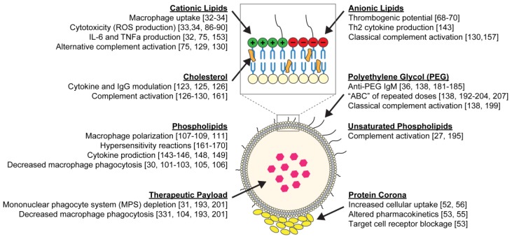
Liposomes hold great potential as gene and drug delivery vehicles due to their biocompatibility and modular properties, coupled with the major advantage of attenuating the risk of systemic toxicity from the encapsulated therapeutic agent. Decades of research have been dedicated to studying and optimizing liposomal formulations for a variety of medical applications, ranging from cancer therapeutics to analgesics. Some effort has also been made to elucidate the toxicities and immune responses that these drug formulations may elicit. Notably, intravenously injected liposomes can interact with plasma proteins, leading to opsonization, thereby altering the healthy cells they come into contact with during circulation and removal. Additionally, due to the pharmacokinetics of liposomes in circulation, drugs can end up sequestered in organs of the mononuclear phagocyte system, affecting liver and spleen function. Importantly, liposomal agents can also stimulate or suppress the immune system depending on their physiochemical properties, such as size, lipid composition, pegylation, and surface charge. Despite the surge in the clinical use of liposomal agents since 1995, there are still several drawbacks that limit their range of applications. This review presents a focused analysis of these limitations, with an emphasis on toxicity to healthy tissues and unfavorable immune responses, to shed light on key considerations that should be factored into the design and clinical use of liposomal formulations.

Keywords: cancer; gene and drug delivery; immunomodulation; liposomes; toxicity.


Conflict of interest statement

The authors declare no conflict of interest.

Figures

Figure 1
Figure 1
Schematic of a liposome and its common building blocks. The liposomal bilayer is composed of phospholipids (neutral, cationic or anionic) and cholesterol, and the surface can be decorated with polyethylene glycol (PEG). Hydrophilic drugs can be entrapped within the aqueous core, while hydrophobic drugs can be loaded into the lipid bilayer. Once injected into the bloodstream, a protein corona, comprised largely of apolipoproteins, immunoglobulins, and complement proteins, is formed on the liposome surface. The protein corona, which is impacted by the liposomal surface chemistry, governs liposome–cell interactions. The immunological and toxicological effects caused by each liposome component, imparted on cells throughout the body, are summarized throughout the diagram.
Figure 2
Figure 2
Intravenously injected liposomes impact cancer progression in vivo. Liposomal polarization of classically activated M1 tumor-associated macrophages (TAMs) to M2-like TAMs can result in carrier-induced immunosuppression and accelerated tumor development. Compared to vehicle control, empty (placebo) liposomes (A) accelerated tumor growth in a TC-1 subcutaneous mouse model and (B) increased the number of metastatic sites in an orthotopically implanted ID8-VEGF-GFP ovarian carcinoma mouse model on day 36. * P=0.03. (Cited from La-Beck et al., 2019 [107]).
Figure 3
Figure 3
Schematic overview of the complement system highlighting two of the main activation pathways (classical vs. alternative). Previous studies have shown that classical pathway activation is initiated by antibody-liposome binding, pegylated liposomes, and anionic liposomes. The alternative pathway can be initiated by cationic liposomes, liposomes containing more than 40 mol% cholesterol, and liposomes larger than 200 nm in diameter. During the complement cascade, C3b opsonin covalently binds to the surface of the liposome, marking it for removal by the MPS. The released anaphylatoxins (C3a, C4a, and C5a) prompt the activation of leukocytes.
Figure 4
Figure 4
Schematic overview of the events that alter the pharmacokinetics of repeated intravenous (IV) doses of pegylated liposomes. The first dose of pegylated liposomes stimulates the production of anti-PEG IgM antibodies by B cells within the spleen. This leads to the accelerated blood clearance of subsequent doses, during which circulating anti-PEG IgMs bind to the liposomes, initiating classical complement activation and decreasing blood circulation half-life.

References

    1. Torchilin V.P. Recent advances with liposomes as pharmaceutical carriers. Nat. Rev. Drug Discov. 2005;4:145–160. doi: 10.1038/nrd1632. - DOI - PubMed
    1. Akbarzadeh A., Rezaei-Sadabady R., Davaran S., Joo S.W., Zarghami N., Hanifehpour Y., Samiei M., Kouhi M., Nejati-Koshki K. Liposome: Classification, preparation, and applications. Nanoscale Res. Lett. 2013;8:102. doi: 10.1186/1556-276X-8-102. - DOI - PMC - PubMed
    1. Bozzuto G., Molinari A. Liposomes as nanomedical devices. Int. J. Nanomed. 2015;10:975–999. doi: 10.2147/IJN.S68861. - DOI - PMC - PubMed
    1. Szoka F., Jr., Papahadjopoulos D. Procedure for preparation of liposomes with large internal aqueous space and high capture by reverse-phase evaporation. Proc. Natl. Acad. Sci. USA. 1978;75:4194–4198. doi: 10.1073/pnas.75.9.4194. - DOI - PMC - PubMed
    1. Bangham A.D., Horne R.W. Negative staining of phospholipids and their structural MODIFICATION BY surface-active agents as observed in the electron microscope. J. Mol. Biol. 1964;8:660–668. doi: 10.1016/S0022-2836(64)80115-7. - DOI - PubMed

LinkOut - more resources