. 2020 Jan 22;12(2):268.
doi: 10.3390/cancers12020268.

Phenethyl Isothiocyanate Suppresses Stemness in the Chemo- and Radio-Resistant Triple-Negative Breast Cancer Cell Line MDA-MB-231/IR Via Downregulation of Metadherin

Affiliations

Phenethyl Isothiocyanate Suppresses Stemness in the Chemo- and Radio-Resistant Triple-Negative Breast Cancer Cell Line MDA-MB-231/IR Via Downregulation of Metadherin

Yen Thi-Kim Nguyen et al. Cancers (Basel). .

Abstract

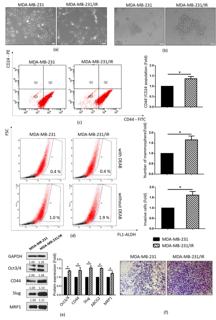
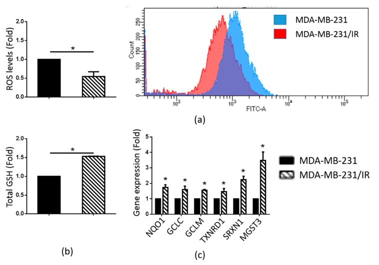
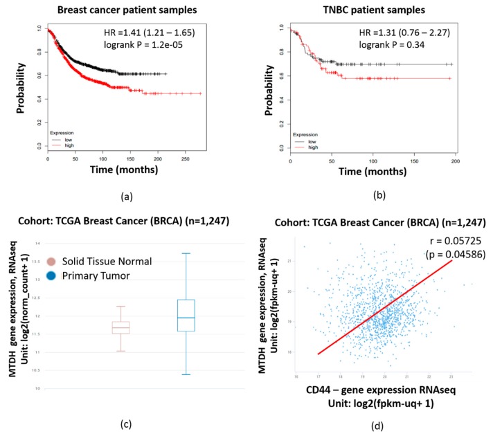
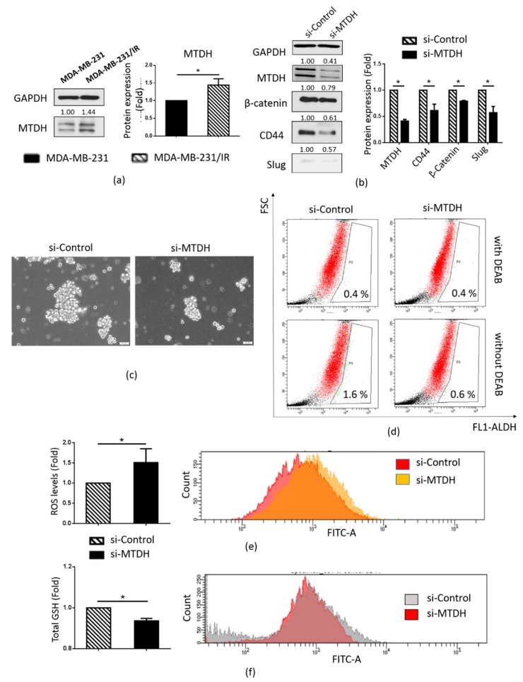
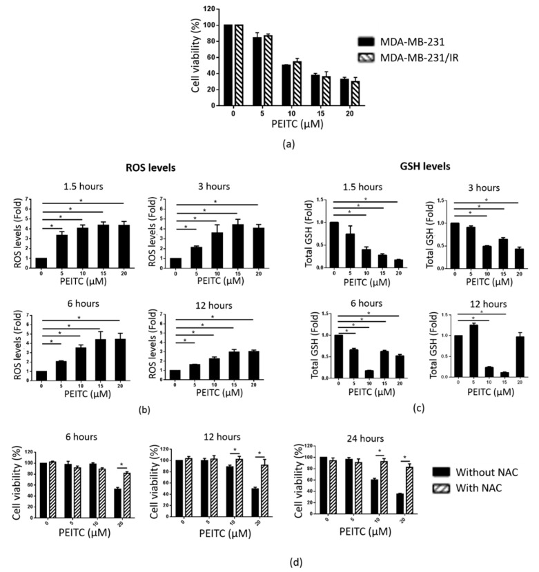
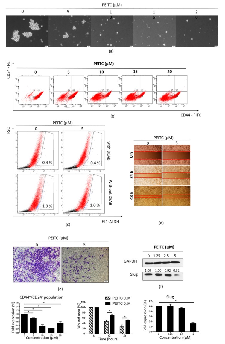
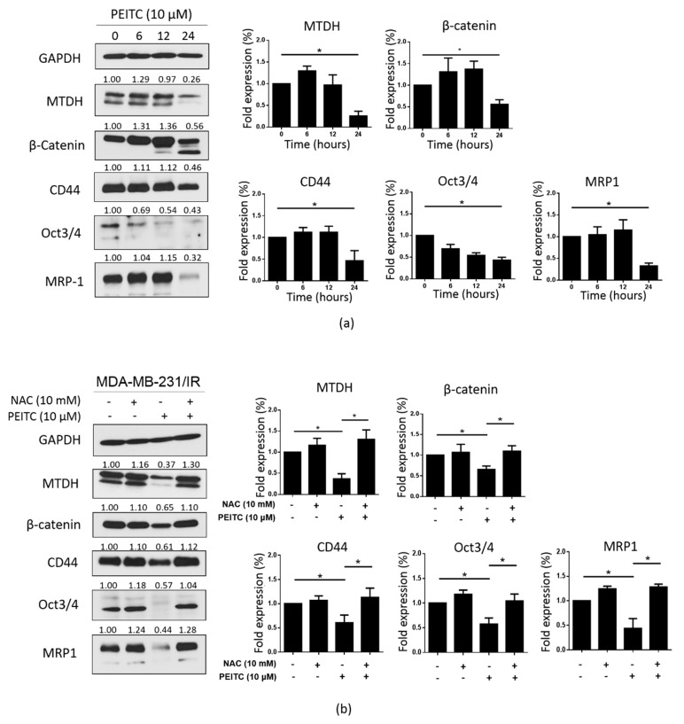
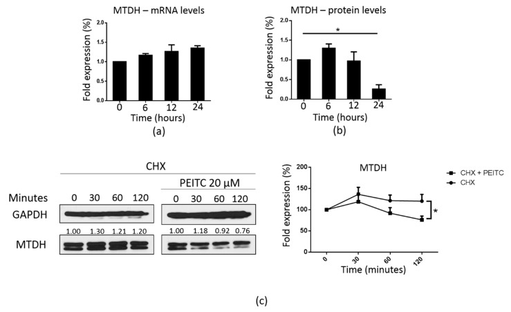
Resistance to chemotherapy and radiation therapy is considered a major therapeutic barrier in breast cancer. Cancer stem cells (CSCs) play a prominent role in chemo and radiotherapy resistance. The established chemo and radio-resistant triple-negative breast cancer (TNBC) cell line MDA-MB-231/IR displays greater CSC characteristics than the parental MDA-MB-231 cells. Escalating evidence demonstrates that metadherin (MTDH) is associated with a number of cancer signaling pathways as well as breast cancer therapy resistance, making it an attractive therapeutic target. Kaplan-Meier plot analysis revealed a correlation between higher levels of MTDH and shorter lifetimes in breast cancer and TNBC patients. Moreover, there was a positive correlation between the MTDH and CD44 expression levels in The Cancer Genome Atlas breast cancer database. We demonstrate that MTDH plays a pivotal role in the regulation of stemness in MDA-MB-231/IR cells. Knockdown of MTDH in MDA-MB-231/IR cells resulted in a reduction in the CSC population, aldehyde dehydrogenase activity, and major CSC markers, including β-catenin, CD44+, and Slug. In addition, MTDH knockdown increased reactive oxygen species (ROS) levels in MDA-MB-231/IR cells. We found that phenethyl isothiocyanate (PEITC), a well-known pro-oxidant phytochemical, suppressed stemness in MDA-MB-231/IR cells through ROS modulation via the downregulation of MTDH. Co-treatment of PEITC and N-Acetylcysteine (a ROS scavenger) caused alterations in PEITC induced cell death and CSC markers. Moreover, PEITC regulated MTDH expression at the post-transcriptional level, which was confirmed using cycloheximide, a protein synthesis inhibitor.

Keywords: cancer stem cells; metadherin; phenethyl isothiocyanate; reactive oxygen species; resistance.


Conflict of interest statement

The authors declare no conflict of interest. The funders had no role in the design of the study; in the collection, analyses, or interpretation of data; in the writing of the manuscript, or in the decision to publish the results.

Figures

Figure 1
Figure 1
Characteristics of radioresistant MDA-MB-231/IR cells. (a) Morphologies of MDA-MB-231 and MDA-MB-231/IR cells cultured as monolayers (100× magnification). (b) MDA-MB-231 and MDA-MB-231/IR cells cultured as mammospheres in complete MammoCult Human Medium for 10 days (100× magnification). (c) CD44+/CD24 population in MDA-MB-231/IR and MDA-MB-231 cells as analyzed by fluorescence-activated cell sorting (FACS). (d) ALDH+ population assessed by the ALDEFLUOR assay kit, with DEAB used as the negative control. (e) Cancer stem cell (CSC) markers were analyzed by Western blotting; * p < 0.05; results are presented as the mean ± standard deviation. (f) Invasion of MDA-MB-231/IR and MDA-MB-231 cells assessed by the Transwell cell invasion assay (100× magnification).
Figure 2
Figure 2
ROS and GSH levels and expression of genes related to antioxidant effects in MDA-MB-231 and MDA-MB-231/IR cells. (a) ROS levels were evaluated after staining with H2DCFDA. (b) Total glutathione levels were measured by the GSH assay. (c) The expression of antioxidant-related genes was analyzed by real-time PCR; * p < 0.05; results are presented as mean ± standard deviation.
Figure 3
Figure 3
Analysis of metadherin (MTDH) expression in breast cancer patients. Correlation between MTDH expression and survival rate in total breast cancer patients (a) and triple negative breast cancer (TNBC) patients (b), as analyzed by Kaplan–Meier plotting. (c) MTDH expression in primary tumor and normal tissue in the The Cancer Genome Atlas (TCGA) breast cancer (BRCA) cohort. (d) Correlation between MTDH and CD44 expression, in the TCGA breast cancer (BRCA) cohort; * p < 0.05; results are presented as mean ± standard deviation.
Figure 4
Figure 4
Role of MTDH in regulating the CSC population in MDA-MB-231/IR cells. (a) Western blot analysis of MTDH in MDA-MB-231 and MDA-MB-231/IR cells. (b) Western blot analysis of MTDH, β-catenin, CD44, and Slug after knockdown by si-MTDH for 48 h. (c) Appearance of mammospheres after siRNA knockdown (100× magnification). (d) ALDH+ population obtained by the ALDEFLUOR assay kit, with DEAB used as a negative control. (e) ROS levels measured by FACS analysis and H2DCFDA staining. (f) The CD44+ population, as assessed by FACS analysis; * p < 0.05; results are presented as mean ± standard deviation.
Figure 5
Figure 5
PEITC reduces cell viability in MDA-MB-231/IR cells by increasing ROS levels. (a) Cell viability was assessed by the MTT assay. (b) ROS levels were analyzed after staining with H2DCFDA. (c) Total GSH levels were measured by a GSH assay. (d) Pre-treatment with NAC (10 mM) helped maintain the viability of MDA-MB-231/IR cells, as assessed by the MTT assay; * p < 0.05; results are presented as mean ± standard deviation.
Figure 6
Figure 6
Effects of PEITC on stemness in MDA-MB-231/IR cells. (a) Mammospheres were cultured in complete MammoCult Human Medium (100× magnification). (b) CD44+/CD24 populations were assessed by FACS analysis after incubation with PEITC (0, 5, 10, 15, or 20 μM) for 24 h. (c) The ALDH+ population was examined using the ALDEFLUOR assay kit, with DEAB used as a negative control; cells were treated with PEITC (0 or 5 μM) for 24 h. (d) Cell migration was determined by the wound healing assay; cells were treated with PEITC (0 or 5 µM) for 24 or 48 h (100× magnification). (e) Invasive cells were stained with crystal violet after treatment with PEITC (0 or 5 µM) for 24 h (100× magnification). (f) Western blot analysis of Slug after incubation of cells with PEITC (0, 1.25, 2.5, or 5 μM) for 24 h; * p < 0.05; results are presented as mean ± standard deviation.
Figure 7
Figure 7
PEITC targets MTDH and CSC markers in MDA-MB-231/IR cells. (a) The levels of MTDH and CSC markers were assessed by Western blotting following PEITC (0, 10 μM) treatment. (b) NAC pre-treatment helped maintain the expression of MTDH and CSC markers; * p < 0.05; results are presented as mean ± standard deviation.
Figure 8
Figure 8
PEITC regulates MTDH post-transcriptionally. (a) Real-time PCR analysis of MTDH following PEITC treatment (10 µM for 6, 12, or 24 h). (b) Western blot analysis was performed to analyze MTDH expression following PEITC exposure (10 µM for 6, 12, or 24 h), similar to Figure 7a. (c) Western blot analysis of the MTDH protein expression following co-treatment with PEITC (20 µM) and cycloheximide (100 μg/mL) for 30, 60, or 120 min; * p < 0.05; results are presented as mean ± standard deviation.

Similar articles

Cited by

References

    1. Foulkes W.D., Smith I.E., Reis-Filho J.S. Triple-negative breast cancer. N. Engl. J. Med. 2010;363:1938–1948. doi: 10.1056/NEJMra1001389. - DOI - PubMed
    1. Langlands F., Horgan K., Dodwell D., Smith L. Breast cancer subtypes: Response to radiotherapy and potential radiosensitisation. Br. J. Radiol. 2013;86:20120601. doi: 10.1259/bjr.20120601. - DOI - PMC - PubMed
    1. Yadav P., Shankar B.S. Radio resistance in breast cancer cells is mediated through TGF-β signalling, hybrid epithelial-mesenchymal phenotype and cancer stem cells. Biomed. Pharmacother. 2019;111:119–130. doi: 10.1016/j.biopha.2018.12.055. - DOI - PubMed
    1. Butti R., Gunasekaran V.P., Kumar T.V., Banerjee P., Kundu G.C. Breast cancer stem cells: Biology and therapeutic implications. Int. J. Biochem. Cell Biol. 2019;107:38–52. doi: 10.1016/j.biocel.2018.12.001. - DOI - PubMed
    1. Diehn M., Cho R.W., Lobo N.A., Kalisky T., Dorie M.J., Kulp A.N., Qian D., Lam J.S., Ailles L.E., Wong M. Association of reactive oxygen species levels and radioresistance in cancer stem cells. Nature. 2009;458:780. doi: 10.1038/nature07733. - DOI - PMC - PubMed