

Applications of Nanocellulose/Nanocarbon Composites: Focus on Biotechnology and Medicine
- PMID: 31979245
- PMCID: PMC7074939
- DOI: 10.3390/nano10020196
Applications of Nanocellulose/Nanocarbon Composites: Focus on Biotechnology and Medicine
Abstract
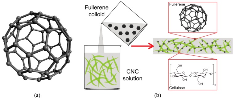
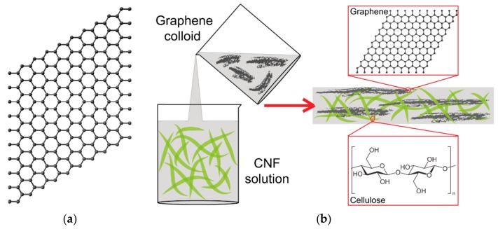
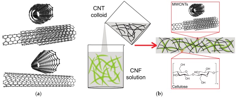
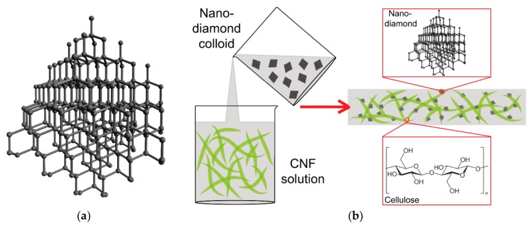
Nanocellulose/nanocarbon composites are newly emerging smart hybrid materials containing cellulose nanoparticles, such as nanofibrils and nanocrystals, and carbon nanoparticles, such as "classical" carbon allotropes (fullerenes, graphene, nanotubes and nanodiamonds), or other carbon nanostructures (carbon nanofibers, carbon quantum dots, activated carbon and carbon black). The nanocellulose component acts as a dispersing agent and homogeneously distributes the carbon nanoparticles in an aqueous environment. Nanocellulose/nanocarbon composites can be prepared with many advantageous properties, such as high mechanical strength, flexibility, stretchability, tunable thermal and electrical conductivity, tunable optical transparency, photodynamic and photothermal activity, nanoporous character and high adsorption capacity. They are therefore promising for a wide range of industrial applications, such as energy generation, storage and conversion, water purification, food packaging, construction of fire retardants and shape memory devices. They also hold great promise for biomedical applications, such as radical scavenging, photodynamic and photothermal therapy of tumors and microbial infections, drug delivery, biosensorics, isolation of various biomolecules, electrical stimulation of damaged tissues (e.g., cardiac, neural), neural and bone tissue engineering, engineering of blood vessels and advanced wound dressing, e.g., with antimicrobial and antitumor activity. However, the potential cytotoxicity and immunogenicity of the composites and their components must also be taken into account.
Keywords: carbon nanotubes; cellulose nanocrystals; diamond nanoparticles; drug delivery; fullerenes; graphene; nanofibrillated cellulose; sensors; tissue engineering; wound dressing.
Conflict of interest statement
The authors declare no conflict of interest. The funding sponsors had no role in the design of the study; in the collection, analyses, or interpretation of data; in the writing of the manuscript, and in the decision to publish the results.
Figures
References
-
- Zhang H., Dou C., Pal L., Hubbe M.A. Review of Electrically Conductive Composites and Films Containing Cellulosic Fibers or Nanocellulose. Bioresources. 2019:14.
-
- Zhang Y.X., Nypelo T., Salas C., Arboleda J., Hoeger I.C., Rojas O.J. Cellulose Nanofibrils: From Strong Materials to Bioactive Surfaces. J Renew Mater. 2013;1:195–211. doi: 10.7569/JRM.2013.634115. - DOI
-
- Lin N., Dufresne A. Nanocellulose in biomedicine: Current status and future prospect. Eur Polym J. 2014;59:302–325. doi: 10.1016/j.eurpolymj.2014.07.025. - DOI
-
- Bhattacharya M., Malinen M.M., Lauren P., Lou Y.R., Kuisma S.W., Kanninen L., Lille M., Corlu A., GuGuen-Guillouzo C., Ikkala O., et al. Nanofibrillar cellulose hydrogel promotes three-dimensional liver cell culture. J Control Release. 2012;164:291–298. doi: 10.1016/j.jconrel.2012.06.039. - DOI - PubMed
Publication types
Grants and funding
LinkOut - more resources
Full Text Sources
