

Ex vivo expansion of regulatory T cells from abdominal aortic aneurysm patients inhibits aneurysm in humanized murine model
- PMID: 31980239
- PMCID: PMC10690961
- DOI: 10.1016/j.jvs.2019.08.285
Ex vivo expansion of regulatory T cells from abdominal aortic aneurysm patients inhibits aneurysm in humanized murine model
Abstract
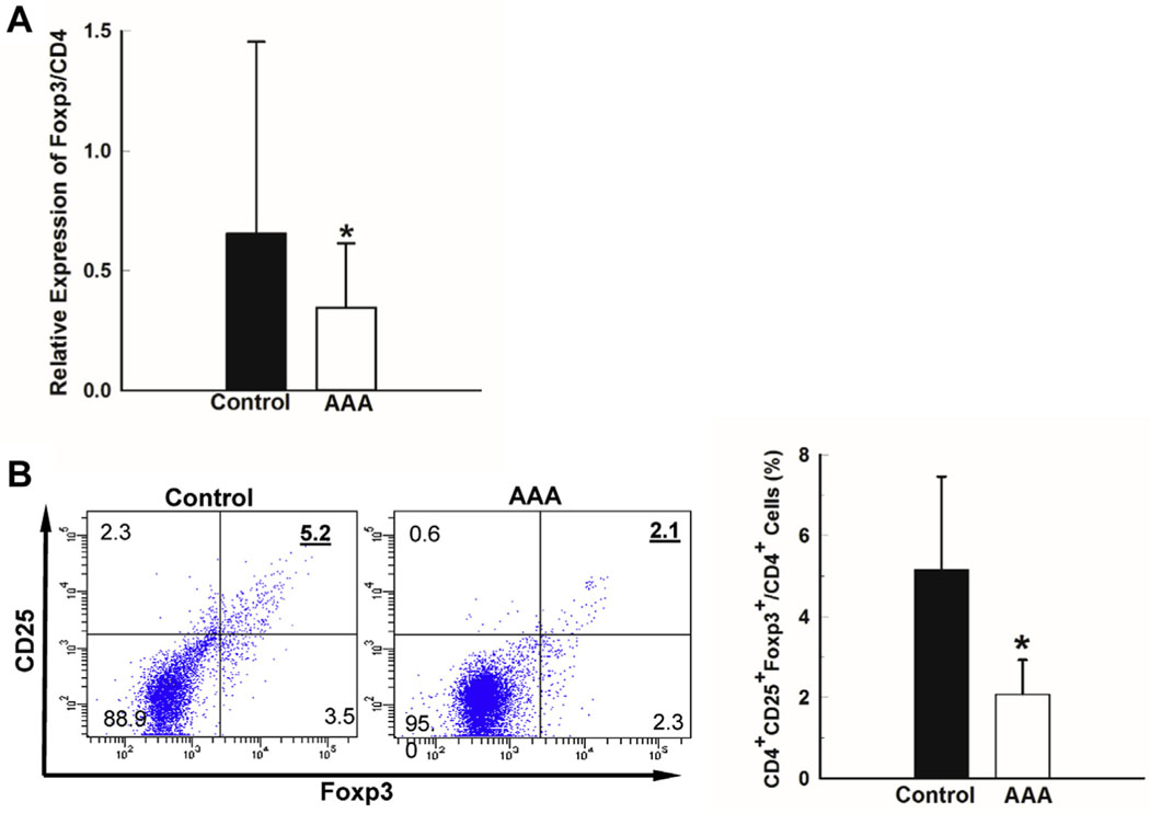
Objective: Abdominal aortic aneurysm (AAA) is a chronic inflammatory disease. Studies of human aneurysm tissue demonstrate dense inflammatory cell infiltrates with CD4+ T cells predominating. Regulatory T cells (Tregs) play an important role in inhibiting pro-inflammatory T cell proliferation, therefore, limiting collateral tissue destruction. The aim of this study was to investigate whether ex vivo augmentation of human Tregs attenuates aneurysm formation in humanized murine model of AAA.
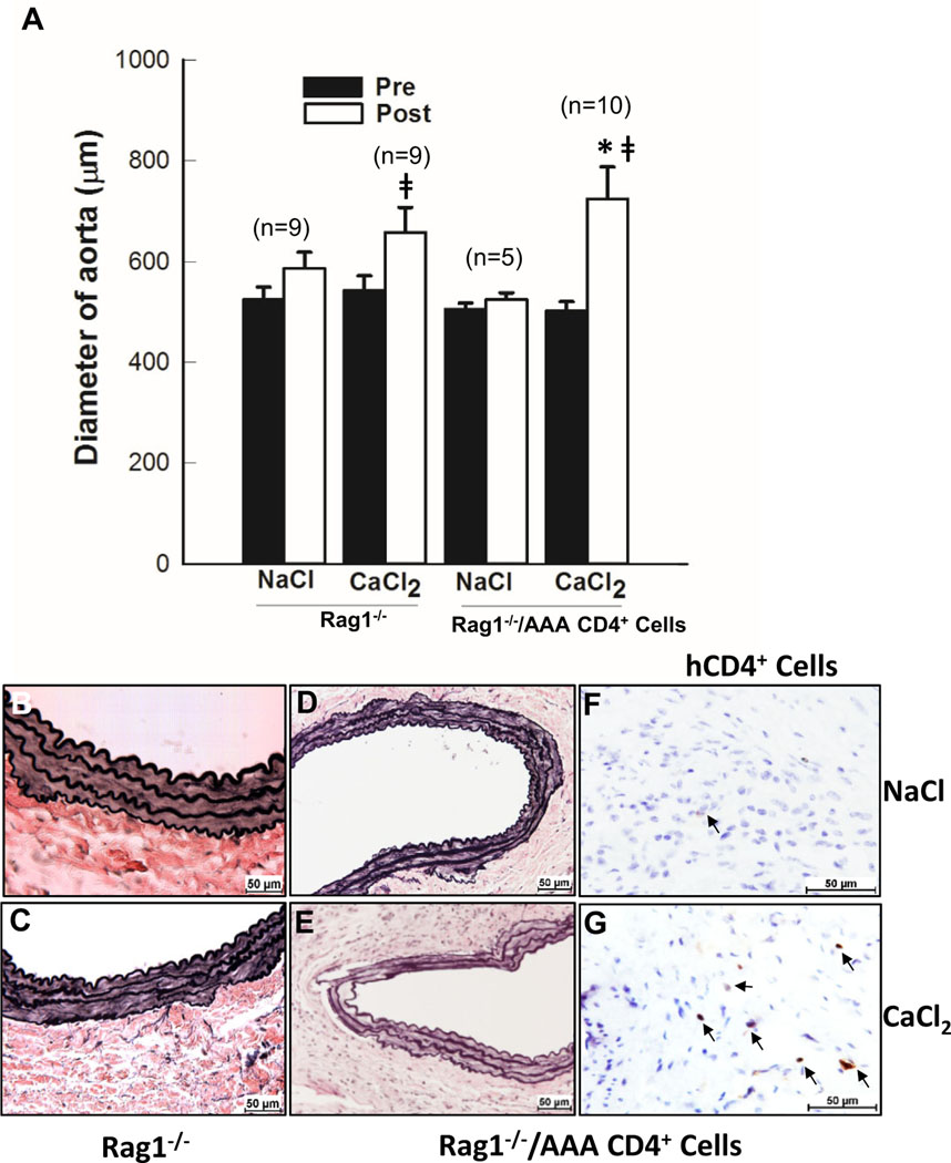
Methods: Circulating Treg population in AAA patients and age- and gender-matched controls were determined by real-time polymerase chain reaction and flow cytometry. To create humanized murine model of AAA, irradiated Rag1-deficient (Rag1-/-) mice, without mature T lymphocytes, at 7 weeks of age were given 5 × 10⁶ of human CD4+ T cells intraperitoneally. Then the mice underwent CaCl2 aneurysm induction. Aortic diameters were measured before and at 6 weeks after aneurysm induction. Aortic tissue was collected for histology and protein extraction. Verhoeff-Van Gieson stain was used for staining elastic fiber. CD4+ T cells in the aortic tissue were detected by immunohistochemical staining.
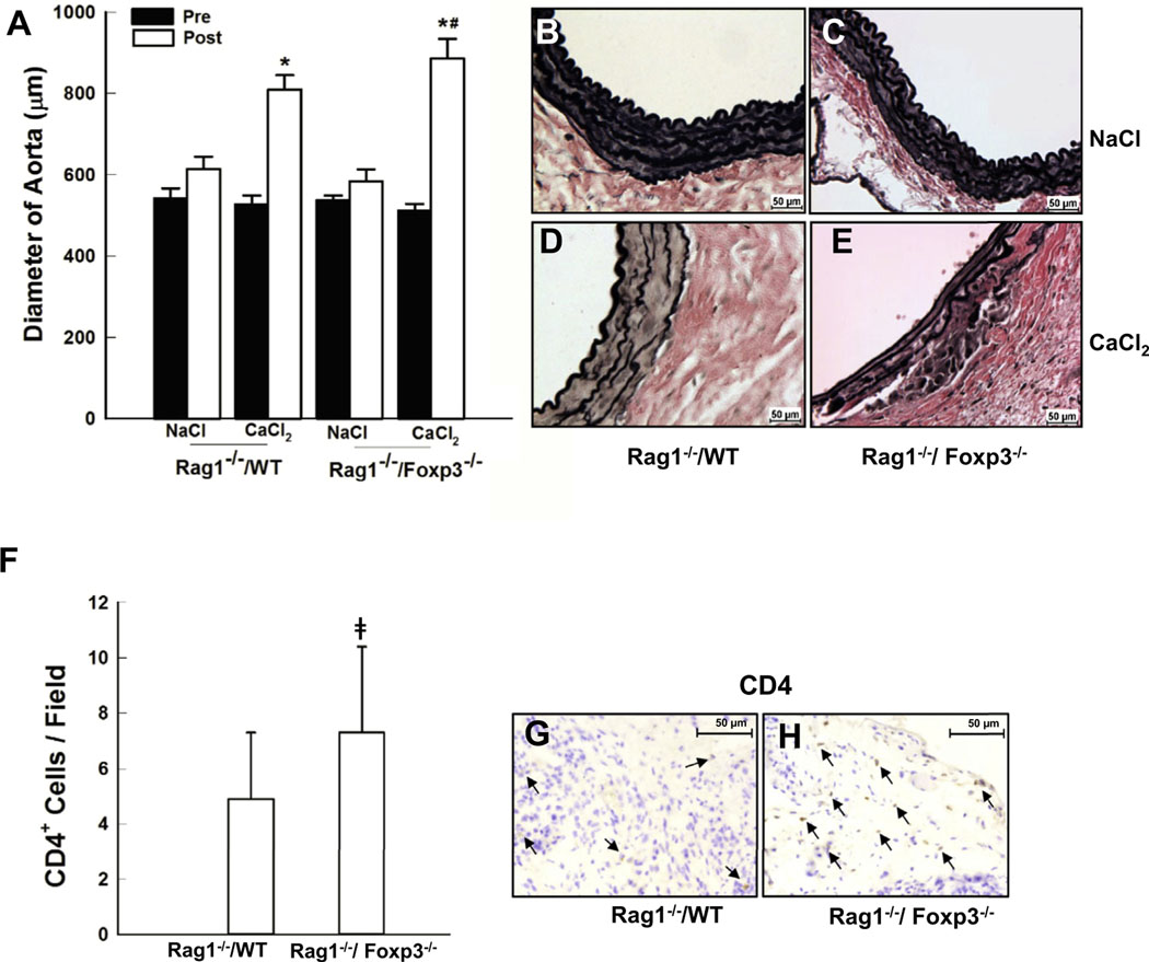
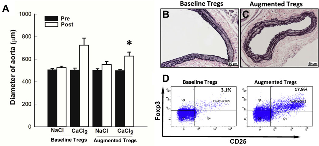
Results: In human peripheral blood mononuclear cells, the proportion of Tregs are decreased in AAA patients compared with matched control patients with significant vascular disease. We first validated the role of Tregs in the CaCl2 model of AAA. To determine the role of human T cells in AAA formation, Rag1-/- mice, resistant to CaCl2-aneurysm induction, were transplanted with human CD4+ T cells. Human CD4+ T cells were able to drive aneurysm formation in Rag1-/- mice. We show that ex vivo augmentation of human Tregs by interleukin-2 resulted in decreased aneurysm progression.
Conclusions: These data suggest that the ex vivo expansion of human Tregs may be a potential therapeutic strategy for inhibiting progression of AAA.
Keywords: Aneurysm; Aorta; Inflammation; Regulatory T cells.
Copyright © 2019 The Authors. Published by Elsevier Inc. All rights reserved.
Conflict of interest statement
Author conflict of interest: none.
Figures
Comment in
-
Recognizing the evolving and beneficial role of regulatory T cells in aneurysm growth.J Vasc Surg. 2020 Sep;72(3):1097. doi: 10.1016/j.jvs.2019.09.045. J Vasc Surg. 2020. PMID: 32829766 No abstract available.
References
-
- Sakalihasan N, Limet R, Defawe OD. Abdominal aortic aneurysm. Lancet 2005;365:1577–89. - PubMed
-
- Duftner C, Seiler R, Klein-Weigel P, Gobel H, Goldberger C, Ihling C, et al. High prevalence of circulating CD4+CD28- T-cells in patients with small abdominal aortic aneurysms. Arterioscler Thromb Vasc Biol 2005;25:1347–52. - PubMed
-
- Chan WL, Pejnovic N, Hamilton H, Liew TV, Popadic D, Poggi A, et al. Atherosclerotic abdominal aortic aneurysm and the interaction between autologous human plaque-derived vascular smooth muscle cells, type 1 NKT, and helper T cells. Circ Res 2005;96:675–83. - PubMed
-
- Ciavarella C, Alviano F, Gallitto E, Ricci F, Buzzi M, Velati C, et al. Human vascular wall mesenchymal stromal cells contribute to abdominal aortic aneurysm pathogenesis through an impaired immunomodulatory activity and increased levels of matrix metalloproteinase-9. Circ J 2015;79:1460–9. - PubMed
Publication types
MeSH terms
Substances
Grants and funding
LinkOut - more resources
Full Text Sources
Research Materials
