Review
. 2020 Apr:100:74-87.
doi: 10.1016/j.semcdb.2019.12.004. Epub 2020 Jan 22.

Cellular and molecular basis of liver regeneration

Affiliations
Review

Cellular and molecular basis of liver regeneration

Sushant Bangru et al. Semin Cell Dev Biol. 2020 Apr.

Abstract

Recent advances in genetics and genomics have reinvigorated the field of liver regeneration. It is now possible to combine lineage-tracing with genome-wide studies to genetically mark individual liver cells and their progenies and detect precise changes in their genome, transcriptome, and proteome under normal versus regenerative settings. The recent use of single-cell RNA sequencing methodologies in model organisms has, in some ways, transformed our understanding of the cellular and molecular biology of liver regeneration. Here, we review the latest strides in our knowledge of general principles that coordinate regeneration of the liver and reflect on some conflicting evidence and controversies surrounding this topic. We consider the prominent mechanisms that stimulate homeostasis-related vis-à-vis injury-driven regenerative responses, highlight the likely cellular sources/depots that reconstitute the liver following various injuries and discuss the extrinsic and intrinsic signals that direct liver cells to proliferate, de-differentiate, or trans-differentiate while the tissue recovers from acute or chronic damage.

Keywords: Hepatocellular plasticity; Lineage-tracing; Liver injury and repair; Single-cell RNA sequencing; Transcriptional and post-transcriptional gene regulation.


Conflict of interest statement

Declaration of Competing Interest The authors declare no conflict of interest.

Figures

Figure 1.
Figure 1.. Regenerative response of the liver to diverse injuries.
(A) Schematic of the hepatic lobule within an adult liver. Hepatocyte subpopulations with regenerative roles are differentially colored based on population-specific marker genes. (B) Homeostatic regeneration takes place due to normal hepatocyte turnover with aging. Axin2+ cells drive regeneration by expanding towards PV, while the Mfsd2a+ population shrinks over time, and TERTHigh and HybHPs remain constant. (C) In acute injury specifically targeting CV regions (e.g. CCl4), TERTHigh and Mfsd2a+ hepatocyte populations expand towards central vein. While Mfsd2a+ cells expand radially outward, TERTHigh hepatocytes divide asymmetrically producing daughter cells with either TERTHigh or TERTLow signatures. (D) In chronic CV injury, Mfsd2a+ cells expand radially, with their progenies repopulating the CV regions, with significant contributions from TERTHigh hepatocytes as well. (E) During portal vein specific chronic injuries, Sox9+ hybrid (HybHPs) and Mfsd2a+ hepatocyte populations are depleted, with TERTHigh hepatocytes providing a major source for new hepatocytes. (F) Schematic of the liver lobule with relative placement of hepatocytes along porto-central axis based on ploidy states. Lower ploidy states (2n, 4n) dominate near PV and CV, whereas higher ploidy states are present primarily within the core of the lobule. Under regenerative conditions such as after 2/3rd partial hepatectomy (PHx), diploid hepatocytes exhibit higher proliferation rates compared to polyploid hepatocytes. CV: Central vein, PV: portal vein & PA: portal artery.
Figure 2.
Figure 2.. Extrinsic and intrinsic signals regulating the hepatic regenerative response.
(A) Schematic of circulating factors in the hepatic lobule before and after 2/3rd PHx. The elongated blue cells are endothelial cells, while the brown cells are hepatocytes. Post-PHx there is a decrease in circulating glucose levels, with increase in levels of bile acids, norepinephrine and multiple growth factors. Blood insulin and EGF levels increase after PHx due to secretions by pancreas and Brunner’s glands respectively. Additionally, increase in the rate of blood flow leads to turbulent flow in the hepatic sinusoids. (B) Cell-cell interactions in the form of paracrine and autocrine signals released by the resident liver cells. Non-parenchymal cells are exposed to circulating factors as well as increased blood flow rates leading to release of cytokines after PHx. TNFα and IL-6 released by Kupffer cells primes hepatocytes from G0 to G1 phase. Other growth factors including TGFα, HGF, and EGF are secreted by other resident cells to initiate hepatocyte proliferation. These cytokines prime the hepatocyte which then releases cytokines that serve both paracrine (VEGF, TGFα, PDGF) and autocrine (TGFα, amphiregulin) functions.
Figure 3.
Figure 3.. Signaling pathways coordinating the hepatic regenerative response.
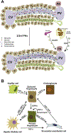
Multiple signaling pathways are stimulated via circulatory factors as well as intrinsic (paracrine and autocrine) cytokines during liver regeneration. After an injury, Kupffer cells are stimulated by circulating LPS as well as the complement system. Kupffer cells play a major role in initiating hepatocyte transition from G0 to G1 phase via induction and release of TNF-α and IL-6. These factors eventually initiate multiple pathways within hepatocytes, including NF-κB, JAK-STAT, PI3K-Akt, and AP1 signaling leading to transcriptional activation. Additionally, Kupffer cells also release the activated Wnt ligand that initiates Wnt/β-Catenin signaling. Hepatocyte growth factor receptor signaling is initiated via both HGF released from ECM by urokinase plasminogen activator (uPA), as well as secreted HGF from hepatic stellate and endothelial cells. EGF receptor signaling is initiated by EGF primarily derived from the Brunner’s gland. EGFR and HGFR signaling feed into the NF-κB and mTOR pathways via PI3K-Akt signaling which can be regulated by miR-382. Sonic hedgehog (Shh) signaling is initiated by Shh secreted from Stellate cells, which in turn inactivates the inhibition of Smo receptor by the Ptch1 receptor. Activation of Smo leads to increased accumulation and localization of Gli to the nucleus and transcriptional response. Also, during regeneration, YAP predominantly localizes to the nucleus interacting with TEAD and β-Catenin proteins to activate transcription of target genes. In the regenerating phase, ESRP2 inhibition leads to depletion of the adult and accumulation of fetal splice isoforms of core Hippo pathway components (e.g., Yap1, Tead1, Nf2, Csnk1d), which suppress Hippo signaling, while promoting nuclear YAP-TEAD interactions and transcriptional activities.
Box Figure.
Box Figure.
(A) Simplified pipeline for identification of new cell types from whole liver suspension as well as analysis of cellular heterogeneity and subtypes after cell sorting using FACS/MACS methods. (B) Isolation of individual cell types in the liver, and ordering of cells using pericentral and periportal dominant genes, to identify zonation patterns at a genome-wide scale. (C) Isolation of specific liver cell subtypes based on marker genes, ploidy status, and pre- or post-liver injury state to identify cellular dynamics with RNA velocity and/or trajectory analysis. Cell-specific trajectories can further resolve the differential transcriptome dynamics between various hepatocyte subtypes.

References

    1. Tanaka EM & Reddien PW The cellular basis for animal regeneration. Dev. Cell 21, 172–185 (2011). - PMC - PubMed
    1. Michalopoulos GK & DeFrances MC Liver regeneration. Science 276, 60–66 (1997). - PubMed
    1. Si-Tayeb K, Lemaigre FP & Duncan SA Organogenesis and development of the liver. Dev. Cell 18, 175–189 (2010). - PubMed
    1. Michalopoulos GK Liver regeneration. J. Cell. Physiol 213, 286–300 (2007). - PMC - PubMed
    1. Vilarinho S & Lifton RP Liver transplantation: from inception to clinical practice. Cell 150, 1096–1099 (2012). - PubMed

Publication types

LinkOut - more resources