. 2020 Feb 11;117(6):2938-2947.
doi: 10.1073/pnas.1915010117. Epub 2020 Jan 24.

Conformational changes upon gating of KirBac1.1 into an open-activated state revealed by solid-state NMR and functional assays

Affiliations

Conformational changes upon gating of KirBac1.1 into an open-activated state revealed by solid-state NMR and functional assays

Reza Amani et al. Proc Natl Acad Sci U S A. .

Abstract

The conformational changes required for activation and K+ conduction in inward-rectifier K+ (Kir) channels are still debated. These structural changes are brought about by lipid binding. It is unclear how this process relates to fast gating or if the intracellular and extracellular regions of the protein are coupled. Here, we examine the structural details of KirBac1.1 reconstituted into both POPC and an activating lipid mixture of 3:2 POPC:POPG (wt/wt). KirBac1.1 is a prokaryotic Kir channel that shares homology with human Kir channels. We establish that KirBac1.1 is in a constitutively active state in POPC:POPG bilayers through the use of real-time fluorescence quenching assays and Förster resonance energy transfer (FRET) distance measurements. Multidimensional solid-state NMR (SSNMR) spectroscopy experiments reveal two different conformers within the transmembrane regions of the protein in this activating lipid environment, which are distinct from the conformation of the channel in POPC bilayers. The differences between these three distinct channel states highlight conformational changes associated with an open activation gate and suggest a unique allosteric pathway that ties the selectivity filter to the activation gate through interactions between both transmembrane helices, the turret, selectivity filter loop, and the pore helix. We also identify specific residues involved in this conformational exchange that are highly conserved among human Kir channels.

Keywords: allostery; lipid activation; membrane protein; potassium channel; solid-state NMR.


Conflict of interest statement

The authors declare no competing interest.

Figures
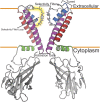
Fig. 1.
Fig. 1.
Anatomy of KirBac1.1. The transmembrane portion of each monomer of KirBac1.1 is composed of four helices: slide helix (green), transmembrane helix 1 (red), pore helix (blue), and transmembrane helix 2 (magenta). The activation gate, most importantly F146, is located at the base of the inner membrane leaflet. The Kir domain, a β-sheet–rich gating bundle, protrudes into the cell cytoplasm. Regions near the top of this domain interact with anionic lipids to actuate channel gating.
Fig. 2.
Fig. 2.
K+ flux assays confirm active channels, and FRET reveals gating motions in the C terminus upon activation. (A) Fluorescent monitoring of K+ flux through the channel allows for real-time determination of the rate of flux. (B) Schematic outline of the K+ fluorescence assay for rate determination. (C) Time-course FRET measurements from which Fmax and F0 measurements were obtained. Alexa Fluor-546 emission was monitored until we observed a maximum plateau. Black, POPC:POPG with A/D-labeled KirBac1.1; blue, POPC:POPG: 1.25% phosphatidylinositol 4,5-bisphosphate (PIP2) labeled with A/D-labeled KirBac1.1; green, POPC:POPG: 3% PIP2 labeled with A/D-labeled KirBac1.1; red, POPC:POPG: 5% PIP2 labeled with A/D-labeled KirBac1.1, nonlabeled; magenta, POPC:POPG: nonlabeled (data are plotted behind cyan curve); cyan, POPC:POPG:PIP2 nonlabeled. Labeled (n = 6) and unlabeled (n = 3) samples are reported as mean ± SEM. (Inset) Chemical structure of PIP2 (D) changes in apparent FRET (mean ± SE) upon different amounts of PIP2 in liposome. **P < 0.05. (E) Cartoon of KirBac1.1 C-terminal domain motion previously reported (2) to be tied to K+ conductance, with the red mark denoting the location of G249 in the crystal structure.
Fig. 3.
Fig. 3.
CSPs of glycine and glycine-adjacent residues observed in the transmembrane region of KirBac1.1 in POPC:POPG membranes. (AD) CSPs observed in the CANcoCA 3D spectra exemplifying the sensitivity to backbone rearrangements. All depicted chemical shift correlations are unique, and the perturbations cannot result from misassignment or ambiguity. (F) Chemical shift perturbations of the 15N backbone residues mapped onto KirBac1.1 (G) The 13Cα chemical shift perturbations. These CSPs indicate an increase in α-helical character of the dominant conductive state, which is quite striking at the top of the pore helix. (H) Absolute value of the sum of all CSPs indicating the global differences between the active and inactive states of the protein.
Fig. 4.
Fig. 4.
Chemical shift perturbations (CSPs) between KirBac1.1 reconstituted in a zwitterionic POPC bilayer (green) and an anionic 3:2 POPC:POPG bilayer (red). (AE) Representative assignment slices of CANcoCA 3D experiment showing (A) structural changes to H117–P118 at the top of the SFL, (B) G85–L84 at the top of TM1 showing reduced α-helical character, (C) significant changes in A109–L108 at the bottom of the selectivity filter, and (D) S59–V58 showing structural changes as a result of slide helix interactions with anionic lipids. (E) T120 does not respond to the lipid environment. Changes in the 15N (F) and 13Cα (G) chemical shifts mapped onto the structure of KirBac1.1.
Fig. 5.
Fig. 5.
Torsion angles of KirBac1.1 predicted by TALOS-N software and torsion angles from the KirBac1.1 crystal structure (PDB ID code 1P7B). (A) Residue-averaged B-factor per residue of the assigned portion of KirBac1.1 crystal structure. (B and C) TALOS-N predicted dihedral angles of KirBac1.1 in 3:2 POPC:POPG lipid bilayer of the major conformer (red) and the minor conformer (blue) and zwitterionic bilayer (green) versus the crystallographic dihedral angles (black).
Fig. 6.
Fig. 6.
Cartoon of the proposed allosteric mechanism for KirBac1.1’s gating/inactivation hypothesis and sequential alignment of KirBac1.1 to human Kir channels. (A) Structural changes in KirBac1.1’s fold resulting from anionic lipid binding. Opening the activation gate introduces slow transitions between open-conductive and open-nonconductive states of the channel. These states may be related to fast gating and rectification phenomena. (B) Sequential alignment of KirBac1.1 illustrating conserved homology between KirBac1.1 and human Kir channels. Circles denote the TM1 and TM2 hinge residues. Stars denote the residues potentially responsible for stabilizing rotation of the pore helix through hydrogen bonding. Triangles show residues at the start of the pore helix that are implicated in rotation of the pore helix. Rectangles are residues at the bottom of the pore helix that may move to reestablish the S4 site.

References

    1. Clarke O. B., et al. , Domain reorientation and rotation of an intracellular assembly regulate conduction in Kir potassium channels. Cell 141, 1018–1029 (2010). - PubMed
    1. Wang S., Lee S. J., Heyman S., Enkvetchakul D., Nichols C. G., Structural rearrangements underlying ligand-gating in Kir channels. Nat. Commun. 3, 617 (2012). - PMC - PubMed
    1. Wang S., Vafabakhsh R., Borschel W. F., Ha T., Nichols C. G., Structural dynamics of potassium-channel gating revealed by single-molecule FRET. Nat. Struct. Mol. Biol. 23, 31–36 (2016). - PMC - PubMed
    1. Wang S., et al. , Potassium channel selectivity filter dynamics revealed by single-molecule FRET. Nat. Chem. Biol. 15, 377–383 (2019). - PMC - PubMed
    1. Coyote-Maestas W., He Y., Myers C. L., Schmidt D., Domain insertion permissibility-guided engineering of allostery in ion channels. Nat. Commun. 10, 290 (2019). - PMC - PubMed

Publication types

MeSH terms

Substances

LinkOut - more resources