Review
. 2020 Feb;31(2):241-256.
doi: 10.1681/ASN.2019050515. Epub 2020 Jan 24.

CFHR Gene Variations Provide Insights in the Pathogenesis of the Kidney Diseases Atypical Hemolytic Uremic Syndrome and C3 Glomerulopathy

Affiliations
Review

CFHR Gene Variations Provide Insights in the Pathogenesis of the Kidney Diseases Atypical Hemolytic Uremic Syndrome and C3 Glomerulopathy

Peter F Zipfel et al. J Am Soc Nephrol. 2020 Feb.

Abstract

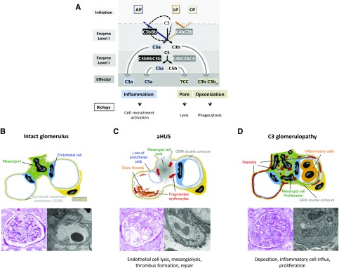
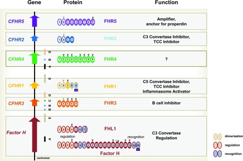
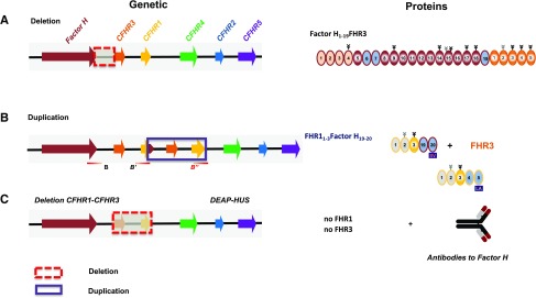
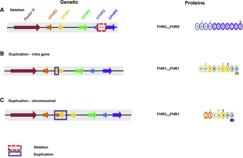
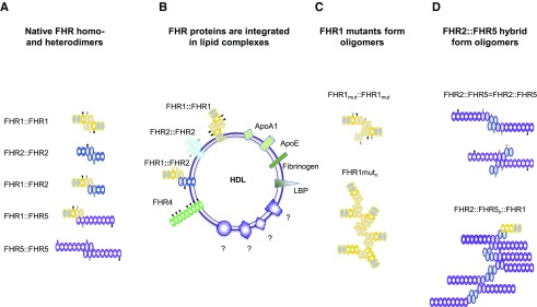
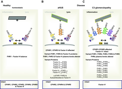
Sequence and copy number variations in the human CFHR-Factor H gene cluster comprising the complement genes CFHR1, CFHR2, CFHR3, CFHR4, CFHR5, and Factor H are linked to the human kidney diseases atypical hemolytic uremic syndrome (aHUS) and C3 glomerulopathy. Distinct genetic and chromosomal alterations, deletions, or duplications generate hybrid or mutant CFHR genes, as well as hybrid CFHR-Factor H genes, and alter the FHR and Factor H plasma repertoire. A clear association between the genetic modifications and the pathologic outcome is emerging: CFHR1, CFHR3, and Factor H gene alterations combined with intact CFHR2, CFHR4, and CFHR5 genes are reported in atypical hemolytic uremic syndrome. But alterations in each of the five CFHR genes in the context of an intact Factor H gene are described in C3 glomerulopathy. These genetic modifications influence complement function and the interplay of the five FHR proteins with each other and with Factor H. Understanding how mutant or hybrid FHR proteins, Factor H::FHR hybrid proteins, and altered Factor H, FHR plasma profiles cause pathology is of high interest for diagnosis and therapy.

Keywords: Complement Factor H related; complement; glomerular disease; glomerulonephritis; hemolytic uremic syndrome.


Figures

Figure 1.
Figure 1.
Overview of complement activation and effector pathways and morphologic changes in aHUS and C3 glomerulopathy. (A) Complement activation occurs via three pathways, the alternative pathway (AP), the lectin pathway (LP), and the classic pathway (CP), which are initiated on surfaces. The type of surface influences activation and the regulator repertoire decides on cascade progression or inhibition. The LP and CP are activated on surfaces by specific carbohydrate moieties (LP), by surface-deposited components (e.g., C reactive protein, Pentraxin), and by IgGs (CP). A repertoire of regulators controls cascade progression in the fluid phase and on surfaces. The three pathways form specific surface-bound convertases; the AP results in the generation of AP-C3 convertase and the LP/CP trigger formation of the CP C3 convertase. The general role of both C3 convertases is to cleave the abundant plasma protein C3 (concentration 1000–1500 μg/ml) into the anaphylatoxin C3a and the opsonic C3b. The enzymatic response on the first enzymatic levels is frequently enhanced by the potent self-amplifying amplification loop. When activation proceeds the C3 convertases attach an additional C3b fragment, form the C3bBbC3b complex, changes substrate specificity and form a C5 convertase. C5 convertases of the AP and of the LP/CP pathways exist. The major role of the C5 convertase is to cleave C5 (plasma concentration 350 μg/ml) into the powerful anaphylatoxin C5a and to generate a surface-binding C5b. C5b subsequently initiates the TCC, which forms lytic pores. (B) Structure of an intact glomerulus. Top: Schematic view of mesangial area (green color) in the filtration unit with endothelial cells (blue), GBM (gray), and podocytes (yellow color). Bottom left: PAS staining of an intact pretransplant kidney. Right: Electron microscopic analysis of a pretransplant kidney with intact glomerular structure. (C) Glomerular changes in aHUS. Top: Schematic presentation of hypocellularity due to cell lysis and loss of endothelial and mesangial cells. Bottom left: PAS staining. Right: EM imaging reveals a pattern of thrombotic microangiopathy. (D) C3 glomerulopathy showing a proliferative pattern with hypercellularity and influx of infiltrating cells. Top: Schematic view of mesangial proliferation and thickening of GBM in MPGN (right glomerulus) and in DDD (left glomerulus). Bottom left: Glomerular changes revealed by PAS staining. Right: EM image showing thickening of the GBM and double contour formation.
Figure 2.
Figure 2.
The human Factor HCFHR gene cluster and the FHR, Factor H protein family. The human Factor H–CFHR gene cluster includes the Factor H gene and the five CFHR genes, which are located on chromosome 1q32. Left side: The CFHR genes are positioned downstream of the Factor H gene (bottom) and are arranged in the order CFHR3, CFHR1, CFHR4, CFHR2, and CFHR5. The human CFHR gene cluster includes interspersed duplicated regions, or segmental duplication elements, which have a high sequence identity and are shown as colored bars below the gene structure. Middle segment: Proteins. Domain organization of the secreted FHR proteins and of Factor H gene products FHL1 and Factor H. Each SCR domain is indicated and the domains of each protein are consecutively numbered. Attached carbohydrate side chains are shown above by the tree-like structure. Constitutively attached carbohydrate side chains have black lines, and facultatively attached carbohydrates have gray lines. The N-terminal multimerization domains of FHR1, FHR2, and FHR5 represented by two SCRs are filled with dashed lines. The C-terminal surface-binding regions of FHR1 and the surface-binding regions of Factor H and FHL1 are shown with a stippled pattern. FHR1: L290A296 indicates the two FHR1-specific residues, which differ from that of Factor H. Bottom: The human Factor H gene encodes two fluid-phase C3 convertase inhibitors, FHL1 and Factor H. The complement regulatory region is located in SCRs 1–4, is shown by the orange patterns, and is shared by FHL1 and Factor H. The C-terminal recognition regions, i.e., SCRs 6–7 (FHL1/Factor H) and SCRs 19–20 (Factor H), are shown by blue patterns. The Factor H–specific two amino acids S1191V1196 in the most C-terminal recognition region are shown in the box below SCR 20. FHL1 has a unique six–amino-acid extension that follows SCR 7. Right side: Identified major functions of each FHR protein and of Factor H and FHL1.
Figure 3.
Figure 3.
CFHR gene variations in aHUS and aHUS-associated FHR protein variants. (A) Deletion of chromosomal segments: Factor H::CFHR3 hybrid gene. Genetic deletion generating a Factor H::FHR3 hybrid gene (left side) and the corresponding Factor H::FHR3 hybrid variant are shown using the color code of Figure 2. The deleted chromosomal segment is shown with the box with red, stippled lines. (B) Insertional mutagenesis (duplications) of chromosomal segments generating a CFHR1::Factor H hybrid gene. Genetic duplication (boxes with blue lines) generates an extra CFHR1::Factor H hybrid gene together with an extra CFHR3 allele (left side), and the corresponding FHR1::Factor H hybrid protein is shown. The changes affect the FHR plasma repertoire (see Table 1). (C) In patients with DEAP-HUS, the deletion of CFHR3-CFHR1 is often associated with the formation of autoantibodies that bind to the C terminus of Factor H.
Figure 4.
Figure 4.
CFHR gene variations in C3 glomerulopathy and associated FHR protein variants. (A) Deletion of large chromosomal segments. Deletion of a large chromosomal segment, which includes exons of the CFHR2 gene and the intragenic region spanning to the CFHR5 gene, generates a CFHR2::CFHR5 hybrid gene. This gene encodes an FHR21–2::FHR5 hybrid protein which is shown on the right. (B) Insertional mutation of intragenetic segments: Duplication of CFHR1 gene segments results in an FHR1 mutant protein. Duplication of the first three exons results in a mutant CFHR1 gene. The encoded protein FHR11–2FHR1 has duplicated interaction segments (right panel). (C) Insertional mutageneses of chromosomal segments results in extra hybrid genes. A duplicated extra gene has CFHR3 exons i–iii linked to the last exons of the CFHR1 gene and generates a new CFHR3::CFHR1 hybrid gene that encodes an FHR31–2::FHR1 hybrid protein.
Figure 5.
Figure 5.
FHR proteins form homodimers and heterodimers; FHR hybrid proteins with two multimerization segments form oligomers. (A) The three proteins FHR1, FHR2, and FHR5 form dimers via their N-terminal domains (gray domains). The N-terminal multimerization segments of each FHR protein interact with a second protein and form dimers and even multimers. FHR1 and FHR2 can form heterodimers. (B) FHR proteins are contained in lipid complexes. FHR1, FHR2, and FHR4-A are associated with lipids HDL and LDLD. Lipids include FHR1 and FHR2 together with ApoAI, LPS-binding protein, fibrinogen, and additional so-far-uncharacterized proteins. FHR4-A is present in triglyceride-rich lipid particles, which also include ApoE, ApoAI, ApoE, ApoAIV, ApoB48, and ApoB100. (C) The FHR1 mutants with two interaction sites form larger complexes. Upper panel: A single FHR1::FHR1 mutant with two interaction segments that bind FHR1, FHR2, and FHR5 and allow formation of larger complexes. Oligomerization or branching is disrupted when FHR1, FHR2, or FHR5 with one interaction segment is integrated. (D) The FHR2::FHR5 hybrid forms larger complexes. Upper panel: A single FHR2::FHR5 hybrid has two interaction segments that allow formation of larger complexes. The FHR2::FHR5 hybrid protein can multimerize via the FHR2 (upper constellation) or via the FHR5 multimerization domain with FHR5 (bottom scenario). Oligomerization or branching is disrupted when FHR1 or FHR2 with one interaction segment is attached to the FHR2 domain, or when FHR5 is bound to the FHR5 domain.
Figure 6.
Figure 6.
FHR mutant and hybrid proteins affect complement regulation at target sites. Gene mutations and copy number variations in the human Factor H–CFHR gene cluster in aHUS and C3 glomerulopathy affect protein structure and protein levels in plasma and influence the fate of complement control at target surfaces. (A) Normal homeotic scenario. FHR1 and Factor H with their homologous C-terminal regions bind to surface-deposited C3b at danger sites and to glycosaminoglycans (GAGs). The proteins are shown and the three C-terminal recognition region is presented, and the FHR1-specific LA and the Factor H–specific SV residues in the most C-terminal domains are shown. The balance of FHR1 and Factor H at modified target sites is influenced by the structure of the proteins; by Factor H, FHL1, and FHR plasma levels; by the binding intensities of the proteins to the ligands; and by the density of C3b and the type and composition of GAGs at sites of damage. FHR1 competes with Factor H for surface binding and, in a proper combination, the two regulators adjust and fine-tune local complement action. Thereby, FHR1 initiates inflammasome activation and Factor H dissociates the alternative pathway C3 convertase and inhibits complement progression. (B) Scenarios in aHUS and C3 glomerulopathy. aHUS scenario, left panel: Factor H::FHR3, Factor H::FHR1, and FHR1::Factor H hybrid proteins together with altered plasma levels influence the local FHR1, Factor H balance at a damage surface (left site). C3 glomerulopathy scenario, right panel: The various FHR hybrid and mutant proteins, many of which having two interacting segments and form large oligomeric complexes, influence the local FHR1, Factor H balance at damaged surfaces. Scenarios in aHUS, left panel: Various scenarios in the form of deletions; insertional mutations, including intragene; as well as chromosomal duplications generate CFHR–Factor H or Factor H–CFHR hybrid genes. In aHUS, Factor H and the CFHR1 and CFHR3 genes are affected, but CFHR4, CFHR2, and CFHR5 genes remain intact. Scenarios in C3 glomerulopathy, right panel: Various scenarios in the form of deletions; insertional mutations, including intragene; as well as chromosomal duplications generate CFHR-CFHR hybrid genes. In C3 glomerulopathy all five CFHR genes can be affected, but the Factor H gene remains intact.

References

    1. Zipfel PF, Skerka C, Chen Q, Wiech T, Goodship T, Johnson S, et al. .: The role of complement in C3 glomerulopathy. Mol Immunol 67: 21–30, 2015. - PubMed
    1. Nester CM, Barbour T, de Cordoba SR, Dragon-Durey MA, Fremeaux-Bacchi V, Goodship TH, et al. .: Atypical aHUS: State of the art. Mol Immunol 67: 31–42, 2015. - PubMed
    1. Noris M, Mescia F, Remuzzi G: STEC-HUS, atypical HUS and TTP are all diseases of complement activation. Nat Rev Nephrol 8: 622–633, 2012. - PubMed
    1. Goodship TH, Cook HT, Fakhouri F, Fervenza FC, Frémeaux-Bacchi V, Kavanagh D, et al. .Conference Participants ;: Atypical hemolytic uremic syndrome and C3 glomerulopathy: Conclusions from a “Kidney Disease: Improving Global Outcomes” (KDIGO) controversies conference. Kidney Int 91: 539–551, 2017. - PubMed
    1. Pickering MC, D’Agati VD, Nester CM, Smith RJ, Haas M, Appel GB, et al. .: C3 glomerulopathy: Consensus report. Kidney Int 84: 1079–1089, 2013. - PMC - PubMed

Publication types

MeSH terms

LinkOut - more resources