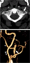
Imaging features of vertebral artery fenestration
- PMID: 31980853
- PMCID: PMC11038258
- DOI: 10.1007/s00234-020-02370-7
Imaging features of vertebral artery fenestration
Abstract
Purpose: Vertebral artery fenestration (VAF) is a rare congenital vascular anomaly which has been associated with intracranial aneurysm. VAF can share some similar imaging features with vertebral artery dissection (VAD), which may confound diagnosis of the latter on CT and MR angiography. The purpose of this investigation is to assess the prevalence of VAF, evaluate its association with other vascular anomalies, and identify imaging features to help in distinguishing VAF and VAD.
Methods: Using keyword search on CTA and MRA head and neck imaging reports from 2010 to 2017, cases of VAF and VAD were retrospectively identified and imaging was reviewed. Imaging features including laterality; vertebral segment; length of affected segment; presence, number, and caliber of lumen(s); and presence of other vascular abnormalities were recorded for all cases and subsequently compared using Pearson's chi-squared test to assess for significant differences between the groups. Patient age, gender, and clinical presentations were also recorded.
Results: Of 64,888 CT and MR angiographic examinations performed, VAF was identified in 67 (0.1%) and VAD in 54 (0.1%) patients. Compared with VADs, VAFs were shorter in length (p < 0.001), wider in luminal diameter (p < 0.001), more likely to occur at the V4 segment (p < 0.01), more likely to have two distinct lumens rather than one (p < 0.01), and less likely to present post-trauma (p < 0.01). Coexisting intracranial aneurysms were identified in 9% of patients with VAF.
Conclusion: VAFs, although rare, can be readily distinguished from VADs on angiographic imaging. Diagnosis of VAF should prompt review for intracranial aneurysm.
Keywords: Angiography; Dissection; Fenestration.
Conflict of interest statement
Figures


References
-
- Tranh-Dinh H, Soo Y, Jayasinghe L (1991) Duplication of the Vertebro-Basilar System. Australas Radiol 35:220–224 - PubMed
-
- Rieger P, Huber G (1983) Fenestration and duplicate origin of the left vertebral artery in angiography. Neuroradiology 25:45–50 - PubMed
-
- Takahasi M, Kawanami H, Watanabe N, Masuoka S (1970) Fenestration of the extracranial vertebral artery. Radiology 96:359–360 - PubMed
MeSH terms
Grants and funding
LinkOut - more resources
Full Text Sources
Medical

