. 2020 Mar;19(3):e13094.
doi: 10.1111/acel.13094. Epub 2020 Jan 25.

Tissue specificity of senescent cell accumulation during physiologic and accelerated aging of mice

Affiliations

Tissue specificity of senescent cell accumulation during physiologic and accelerated aging of mice

Matthew J Yousefzadeh et al. Aging Cell. 2020 Mar.

Abstract

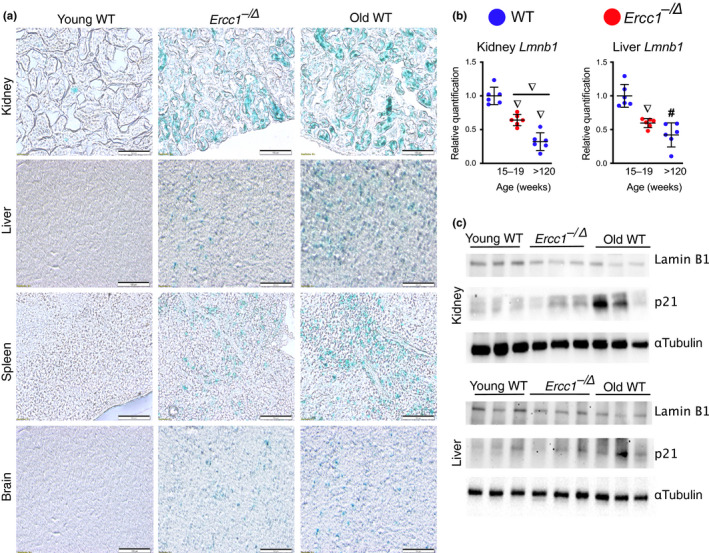
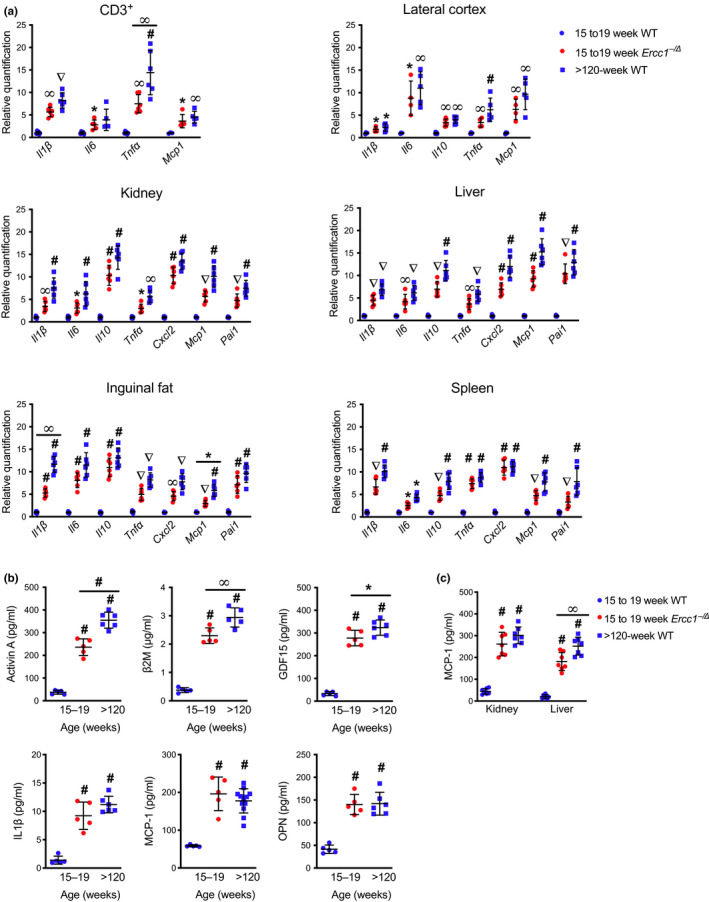
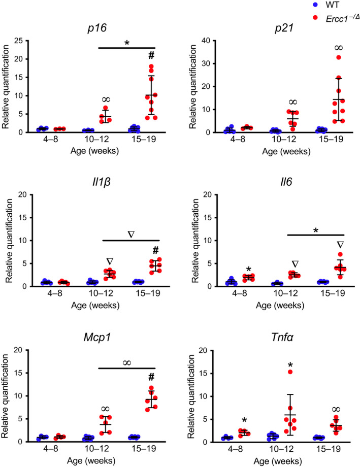
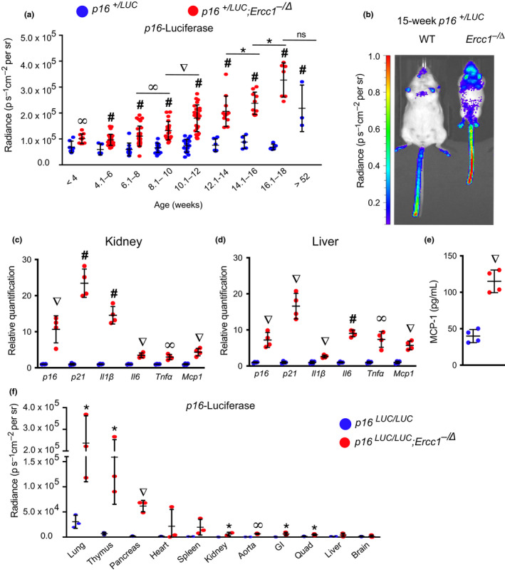
Senescent cells accumulate with age in vertebrates and promote aging largely through their senescence-associated secretory phenotype (SASP). Many types of stress induce senescence, including genotoxic stress. ERCC1-XPF is a DNA repair endonuclease required for multiple DNA repair mechanisms that protect the nuclear genome. Humans or mice with reduced expression of this enzyme age rapidly due to increased levels of spontaneous, genotoxic stress. Here, we asked whether this corresponds to an increased level of senescent cells. p16Ink4a and p21Cip1 mRNA were increased ~15-fold in peripheral lymphocytes from 4- to 5-month-old Ercc1-/∆ and 2.5-year-old wild-type (WT) mice, suggesting that these animals exhibit a similar biological age. p16Ink4a and p21Cip1 mRNA were elevated in 10 of 13 tissues analyzed from 4- to 5-month-old Ercc1-/∆ mice, indicating where endogenous DNA damage drives senescence in vivo. Aged WT mice had similar increases of p16Ink4a and p21Cip1 mRNA in the same 10 tissues as the mutant mice. Senescence-associated β-galactosidase activity and p21Cip1 protein also were increased in tissues of the progeroid and aged mice, while Lamin B1 mRNA and protein levels were diminished. In Ercc1-/Δ mice with a p16Ink4a luciferase reporter, bioluminescence rose steadily with age, particularly in lung, thymus, and pancreas. These data illustrate where senescence occurs with natural and accelerated aging in mice and the relative extent of senescence among tissues. Interestingly, senescence was greater in male mice until the end of life. The similarities between Ercc1-/∆ and aged WT mice support the conclusion that the DNA repair-deficient mice accurately model the age-related accumulation of senescent cells, albeit six-times faster.

Keywords: DNA repair; ERCC1-XPF; aging; cellular senescence; endogenous DNA damage; progeria.


Conflict of interest statement

None declared.

Figures

Figure 1
Figure 1
Expression of p16Ink4a and p21Cip1 mRNA in various murine tissues with aging. (a) Total RNA was isolated from CD3+ T lymphocytes purified from the peripheral blood of 15‐ to 19‐week‐old Ercc1−/∆ (red) mice, age‐matched WT controls, and aged WT (>120‐week‐old) mice (blue) (n = 5–8 per group) by magnetic bead purification using an anti‐CD3 antibody. Expression of the senescence genes, p16Ink4a and p21Cip1, was measured by qPCR. Total RNA was isolated from snap frozen tissues collected from Ercc1−/∆ (red) and WT (blue) mice (n = 3–15 mice per group). Expression of (b) p16Ink4a and (c) p21Cip1 was measured by qPCR using the ∆∆Ct method and normalized to Gapdh expression. Values represent the mean ± SD, one‐way ANOVA with Tukey's test. *p < .05, p < .01, p < .001, # p < .0001
Figure 2
Figure 2
Expression of other senescence markers in WT and progeroid mice. (a) Representative images of fixed frozen samples from 18‐week‐old Ercc1−/∆ mice, age‐matched WT controls, and old WT (>120‐week‐old) mice stained for SA‐βgal activity and imaged at 20X with bright‐field microscopy (scale bar, 100 μm). (b) Total RNA was isolated from snap frozen tissues collected from Ercc1−/∆ (red) and WT (blue) mice (n = 6 mice per group). Expression of the senescence marker LmnB1 was measured by qPCR using the ∆∆Ct method and normalized to Gapdh expression. (c) Lysates from the kidney and liver of 15‐ to 19‐week‐old Ercc1−/∆ mice, age‐matched “young” WT controls and “old” WT (>120‐week‐old) mice were immunoblotted with an anti‐Lamin B1 or ‐p21Cip1 antibodies. α‐Tubulin served as a loading control. Values represent the mean ± SD, one‐way ANOVA with Tukey's test. p < .001, # p < .0001
Figure 3
Figure 3
Expression of senescence‐associated secretory phenotype genes in various murine tissues with aging. (a) Total RNA was isolated from CD3+ T lymphocytes purified from peripheral blood or from snap frozen tissues isolated from 15‐ to 19‐week‐old Ercc1−/∆ mice, age‐matched WT controls, and “old” WT (>120‐week‐old) mice (n = 3–7 per group). Expression of senescence‐associated secretory phenotype (SASP) genes was measured by qPCR using the ∆∆Ct method and normalized to Gapdh expression. (b) Circulating levels of SASP and pro‐geronic factors were quantified in the serum of mice by ELISA (n = 5–19 mice per group). (c) Tissue levels of the SASP factor MCP‐1 were quantified by ELISA (n = 5–12 mice per group). Values represent the mean ± SD, one‐way ANOVA with Tukey's test. *p < .05, p < .01, p < .001, # p < .0001
Figure 4
Figure 4
Progressive increase in expression of senescence markers in Ercc1−/∆ mice. Total RNA was isolated from snap frozen liver collected from Ercc1−/∆ (red) and WT (blue) mice (n = 4–9 mice per group). Senescence (p16Ink4a and p21Cip1) and SASP factor (Il1β, Il6, Mcp1, and Tnfα) expression were measured by qPCR. Relative expression level was quantified using the ∆∆Ct method and normalized to Gapdh expression. Values represent the mean ± SD, two‐way ANOVA, and Tukey's test. *p < .05, p < .01, p < .001, # p < .0001
Figure 5
Figure 5
Longitudinal quantification of p16Ink4a expression. (a) Total body luciferase activity in p16+/LUC;Ercc1 (red) and p16+/LUC (blue) mice with increasing age (n = 9–30 mice per group). (b) Representative images of in vivo luciferase imaging in a 15‐week‐old p16+/LUC;Ercc1 mouse and a p16+/LUC littermate. Total RNA was isolated from (c) kidney and (d) liver collected from 11‐week‐old p16+/LUC;Ercc1 mice (red) and p16+/LUC littermates (blue) (n = 4 mice per group). Senescence (p16Ink4a and p21Cip1) and SASP factor (Il1β, Il6, Mcp1, and Tnfα) expression was measured by qPCR. Relative expression level was quantified using the ∆∆Ct method and normalized to Gapdh expression. (e) Serum levels of the SASP factor MCP‐1 were quantified in 11‐week‐old mice by ELISA. (f) Total luciferase activity in tissues harvested from 17‐week‐old p16LUC/LUC;Ercc1 (red) and p16LUC/LUC (blue) mice (n = 3 per genotype). Values represent the mean ± SD, unpaired two‐tailed Student's t test and two‐way ANOVA with Tukey's test. *p < .05, p < .01, p < .001, # p < .0001

References

    1. Ahmad, A. , Robinson, A. R. , Duensing, A. , van Drunen, E. , Beverloo, H. B. , Weisberg, D. B. , … Niedernhofer, L. J. (2008). ERCC1‐XPF endonuclease facilitates DNA double‐strand break repair. Molecular and Cellular Biology, 28(16), 5082–5092. 10.1128/MCB.00293-08 - DOI - PMC - PubMed
    1. Baker, D. J. , Childs, B. G. , Durik, M. , Wijers, M. E. , Sieben, C. J. , Zhong, J. , … van Deursen, J. M. (2016). Naturally occurring p16(Ink4a)‐positive cells shorten healthy lifespan. Nature, 530(7589), 184–189. 10.1038/nature16932 - DOI - PMC - PubMed
    1. Baker, D. J. , Jeganathan, K. B. , Cameron, J. D. , Thompson, M. , Juneja, S. , Kopecka, A. , … van Deursen, J. M. (2004). BubR1 insufficiency causes early onset of aging‐associated phenotypes and infertility in mice. Nature Genetics, 36(7), 744–749. 10.1038/ng1382 - DOI - PubMed
    1. Burd, C. E. , Sorrentino, J. A. , Clark, K. S. , Darr, D. B. , Krishnamurthy, J. , Deal, A. M. , … Sharpless, N. E. (2013). Monitoring tumorigenesis and senescence in vivo with a p16(INK4a)‐luciferase model. Cell, 152(1–2), 340–351. 10.1016/j.cell.2012.12.010 - DOI - PMC - PubMed
    1. Burtner, C. R. , & Kennedy, B. K. (2010). Progeria syndromes and ageing: What is the connection? Nature Reviews Molecular Cell Biology, 11(8), 567–578. 10.1038/nrm2944 - DOI - PubMed

Publication types

MeSH terms