. 2020 Feb 20;27(2):223-231.e4.
doi: 10.1016/j.chembiol.2020.01.003. Epub 2020 Jan 24.

A Toxic RNA Catalyzes the Cellular Synthesis of Its Own Inhibitor, Shunting It to Endogenous Decay Pathways

Affiliations

A Toxic RNA Catalyzes the Cellular Synthesis of Its Own Inhibitor, Shunting It to Endogenous Decay Pathways

Raphael I Benhamou et al. Cell Chem Biol. .

Abstract

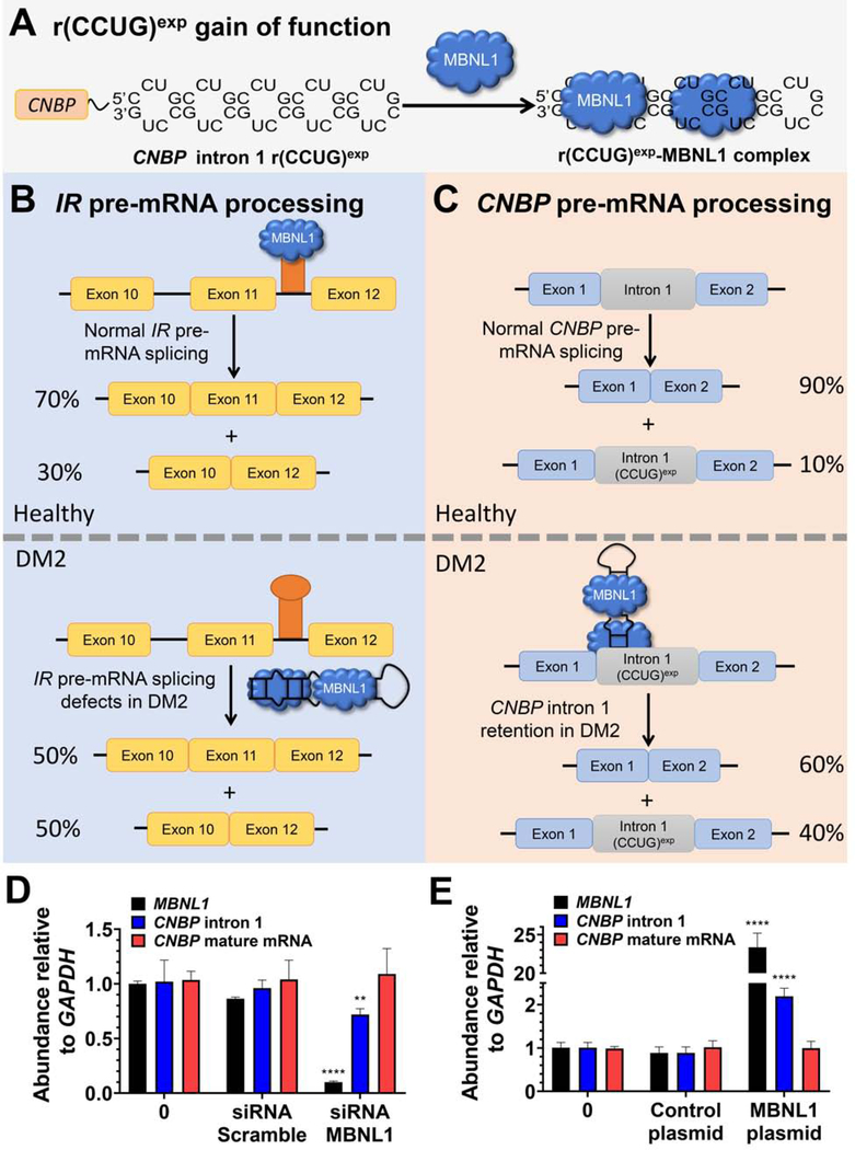
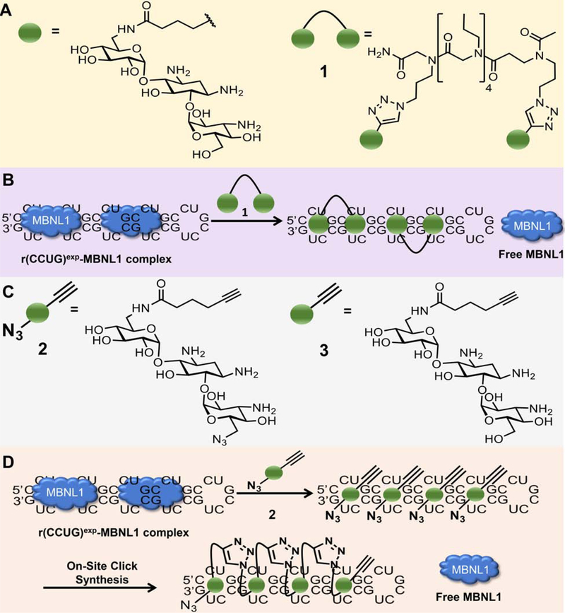
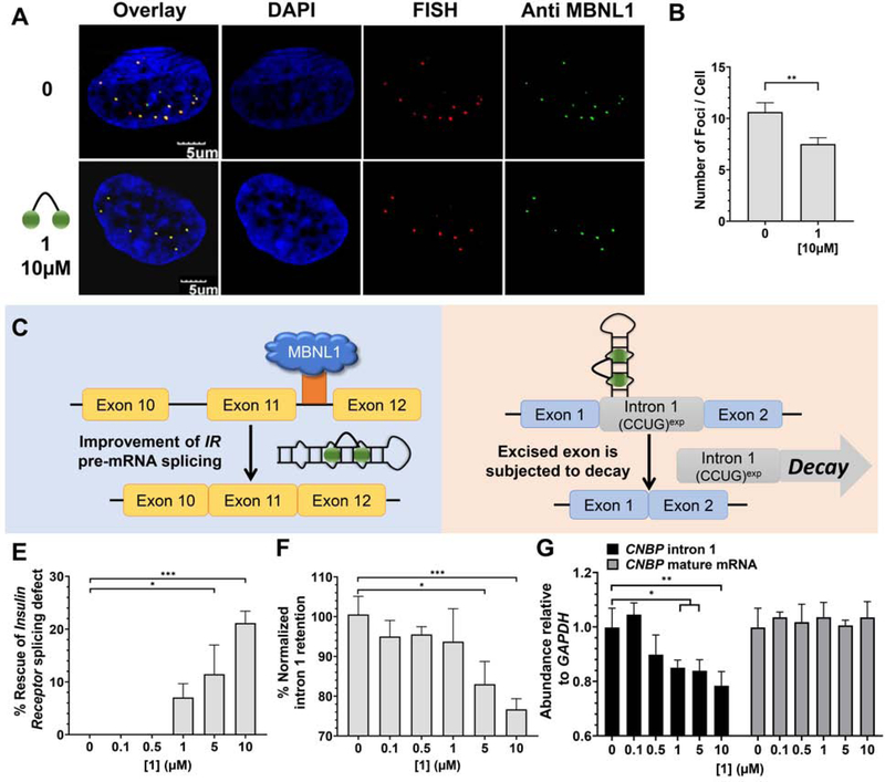
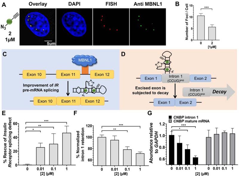
Myotonic dystrophy type 2 (DM2) is a genetically defined disease caused by a toxic expanded repeat of r(CCUG) [r(CCUG)exp], harbored in intron 1 of CCHC-type zinc-finger nucleic acid binding protein (CNBP) pre-mRNA. This r(CCUG)exp causes toxicity via a gain-of-function mechanism, resulting in three pathological hallmarks: aggregation into nuclear foci; sequestration of muscleblind-like-1 (MBNL1) protein, leading to splicing defects; and retention of CNBP intron 1. We studied two types of small molecules with different modes of action, ones that simply bind and ones that are templated by r(CCUG)exp in cells, i.e., the RNA synthesizes its own drug. Indeed, our studies completed in DM2 patient-derived fibroblasts showed that the compounds disrupt the r(CCUG)exp-MBNL1 complex, reduce intron retention, subjecting the liberated intronic r(CCUG)exp to native decay pathways, and rescue other DM2-associated cellular defects. Importantly, this study shows that small molecules can modulate RNA biology by shunting toxic transcripts toward native decay pathways.

Keywords: RNA; chemical biology; click chemistry; drug design; intron retention; medicinal chemistry; microsatellite disease; myotonic dystrophy; nucleic acids; repeat expansion disorder.


Conflict of interest statement

Declaration of Interests M.D.D. is a founder of Expansion Therapeutics, and M.D.D. and E.T.W. are scientific consultants for Expansion Therapeutics.

Figures

Figure 1:
Figure 1:. The RNA that causes DM2, r(CCUG)exp, has multiple modes of toxicity.
(A) r(CCUG)exp in intron 1 of CNBP folds into a hairpin displaying a periodic array of internal loops that sequester MBNL1. Sequestration of MBNL1 by r(CCUG)exp causes (B) pre-mRNA splicing defects, for example increasing IR exon 11 exclusion and (C) aberrant retention of CNBP intron 1. (D) MBNL1 knock-down using siRNA and its effect on relative abundance of MBNL1, CNBP intron 1, and CNBP mature mRNA as measured by RT-qPCR. (E) MBNL1 knock-in using a transfected MBNL1 plasmid and its effect on relative abundance of MBNL1, CNBP intron 1, and CNBP mature mRNA as measured by RT-qPCR. Error bars represent SD. **P < 0.01, ***P < 0.001, ****P < 0.0001, as determined by a one-way ANOVA by comparison to untreated cells (“0”; n = 3). See also Figure S1.
Figure 2:
Figure 2:. Design and biological impact of small molecules targeting r(CCUG)exp.
(A) Structures of a kanamycin derivative (green spheres) that avidly binds the internal loops found in r(CCUG)exp and a dimer composed of two kanamycin binding modules connected via a propylamine peptoid (1). (B) Schematic of 1’s mode of action, binding to r(CCUG)exp and releasing MBNL1. (C) Structures of compounds employed in an on-site drug synthesis approach. Compound 2, a modified kanamycin module, contains alkyne and azide moieties that react upon binding r(CCUG)exp, the catalyst. Compound 3 is a kanamycin module containing only the alkyne component and thus cannot oligomerize. (D) Schematic of on-site click synthesis of 2, catalyzed by binding to r(CCUG)exp, and release MBNL1 to relieve DM2-associated defects.
Figure 3:
Figure 3:. Biological activity of 1 in DM2 patient-derived fibroblasts.
(A) Representative microscopic images for the reduction of nuclear foci by 1, as completed by RNA-FISH and immunohistochemistry using an anti-MBNL1 antibody. (B) Quantification of r(CCUG)exp-MBNL1 foci/nucleus (n = 3 biological replicates, 40 nuclei counted per replicate). (C) Effect of 1 on IR pre-mRNA splicing regulated by MBNL1. (D) Effect of 1 on CNBP pre-mRNA splicing. (E) Effect of 1 on an MBNL1-regulated alternative splicing event, IR exon 11, as determined by RT-PCR; (F) Effect of 1 on aberrant CNBP pre-mRNA splicing caused by r(CCUG)exp, as determined by RT-PCR; that is, retention of intron 1; (G) Effect of 1 on total intron 1 levels and CNBP mature mRNA, as determined by RT-qPCR. Error bars represent SD. *P < 0.5, **P < 0.01, ***P < 0.001, ****P < 0.0001, as determined by a one-way ANOVA (n = 3). See also Figure S2.
Figure 4:
Figure 4:. Biological activity of 2 in DM2 patient-derived fibroxblasts.
(A) Representative microscopic images for the reduction of nuclear foci by 2, as completed by RNA-FISH and immunohistochemistry using an anti-MBNL1 antibody. (B) Quantification of r(CCUG)exp-MBNL1 foci/nucleus (n = 3 biological replicates, 40 nuclei counted per replicate). (C) Effect of 2 on IR pre-mRNA splicing regulated by MBNL1. (D) Effect of 2 on CNBP pre-mRNA splicing. (E) Effect of 2 on an MBNL1- regulated alternative splicing event, IR exon 11, as determined by RT-PCR; (F) Effect of 2 on aberrant CNBP pre-mRNA splicing caused by r(CCUG)exp, as determined by RT-PCR; that is, retention of intron 1; (G) Effect of 2 on total intron 1 levels and CNBP mature mRNA, as determined by RT-qPCR. Error bars represent SD. *P < 0.5, **P < 0.01, ***P < 0.001, as determined by a one-way ANOVA (n = 3). See also Figure S3.

References

    1. Angelbello AJ, Rzuczek SG, McKee KK, Chen JL, Olafson H, Cameron MD, Moss WN, Wang ET, and Disney MD (2019). Precise small-molecule cleavage of an r(CUG) repeat expansion in a myotonic dystrophy mouse model. Proc. Natl. Acad. Sci. U. S. A 116, 7799–7804. - PMC - PubMed
    1. Bernat V, and Disney MD (2015). RNA structures as mediators of neurological diseases and as drug targets. Neuron 87, 28–46. - PMC - PubMed
    1. Chen CZ, Sobczak K, Hoskins J, Southall N, Marugan JJ, Zheng W, Thornton CA, and Austin CP (2012). Two high-throughput screening assays for aberrant RNA-protein interactions in myotonic dystrophy type 1. Anal. Bioanal. Chem 402, 1889–1898. - PMC - PubMed
    1. Childs-Disney JL, Wu M, Pushechnikov A, Aminova O, and Disney MD (2007). A small molecule microarray platform to select RNA internal loop-ligand interactions. ACS Chem. Biol 2, 745–754. - PubMed
    1. Childs-Disney JL, Yildirim I, Park H, Lohman JR, Guan L, Tran T, Sarkar P, Schatz GC, and Disney MD (2014). Structure of the myotonic dystrophy type 2 RNA and designed small molecules that reduce toxicity. ACS Chem. Biol 9, 538–550. - PMC - PubMed

Publication types

MeSH terms