. 2020 Mar 4;28(3):889-900.
doi: 10.1016/j.ymthe.2019.12.014. Epub 2020 Jan 14.

An Optimized Full-Length FLT3/CD3 Bispecific Antibody Demonstrates Potent Anti-leukemia Activity and Reversible Hematological Toxicity

Affiliations

An Optimized Full-Length FLT3/CD3 Bispecific Antibody Demonstrates Potent Anti-leukemia Activity and Reversible Hematological Toxicity

Yik Andy Yeung et al. Mol Ther. .

Abstract

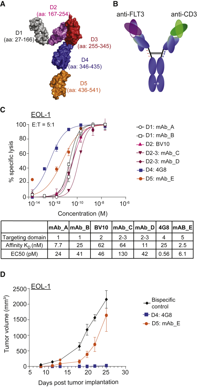
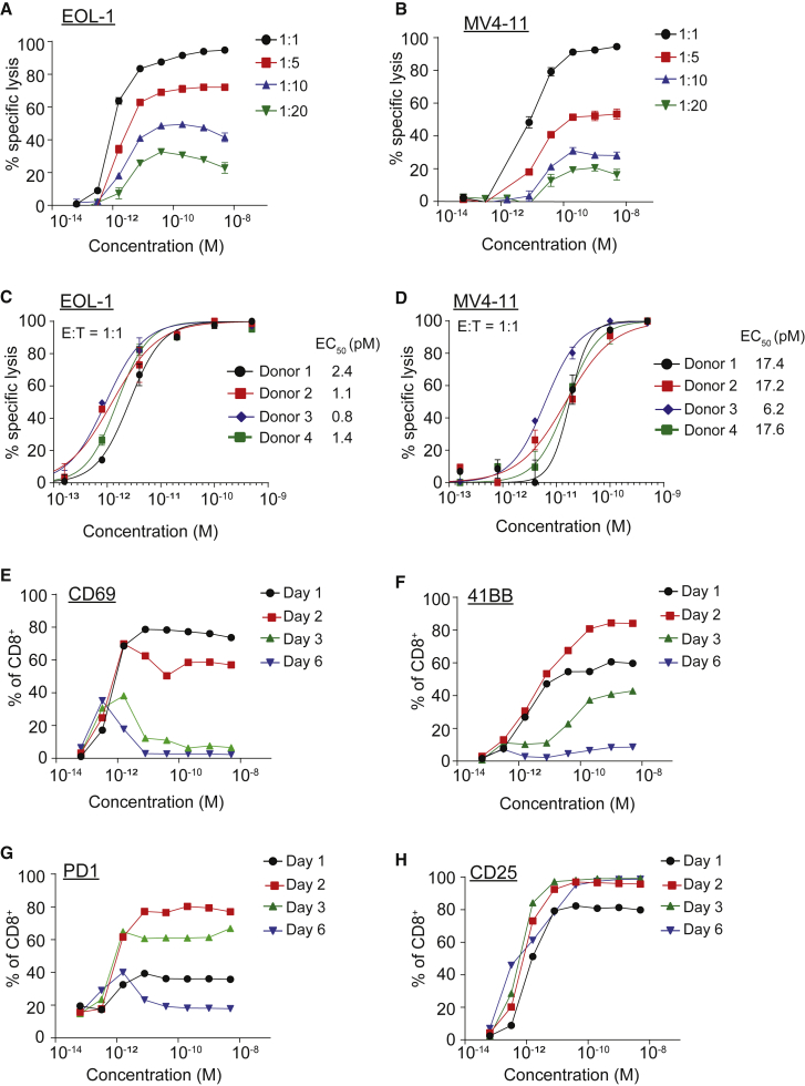
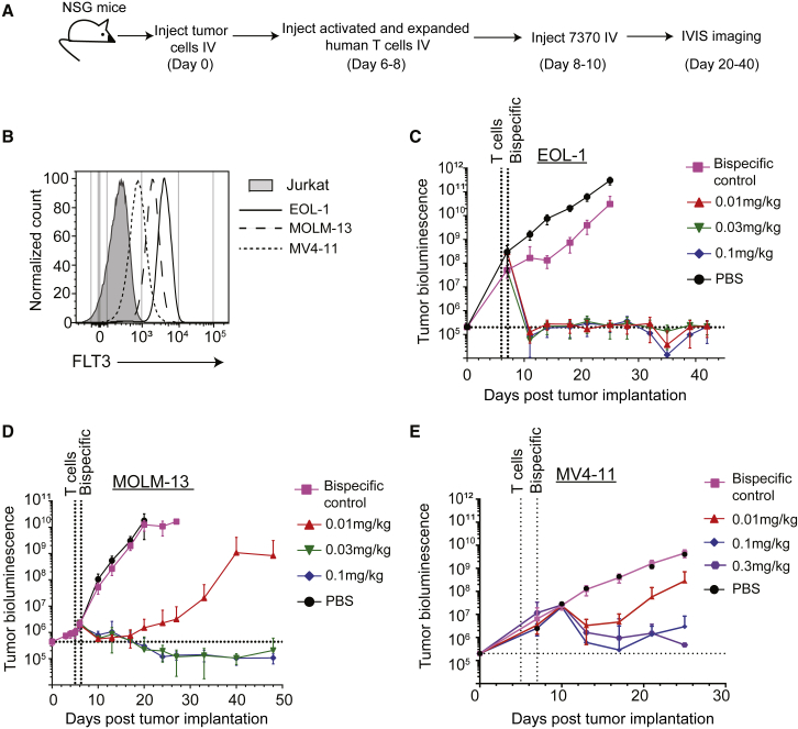
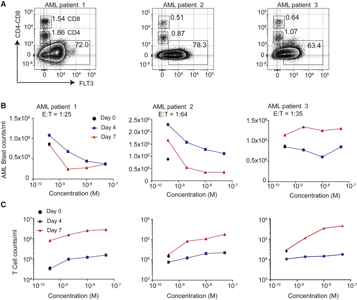
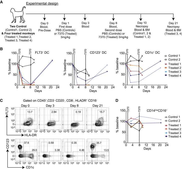
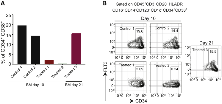
FLT3 (FMS-like tyrosine kinase 3), expressed on the surface of acute myeloid leukemia (AML) blasts, is a promising AML target, given its role in the development and progression of leukemia, and its limited expression in tissues outside the hematopoietic system. Small molecule FLT3 kinase inhibitors have been developed, but despite having clinical efficacy, they are effective only on a subset of patients and associated with high risk of relapse. A durable therapy that can target a wider population of AML patients is needed. Here, we developed an anti-FLT3-CD3 immunoglobulin G (IgG)-based bispecific antibody (7370) with a high affinity for FLT3 and a long half-life, to target FLT3-expressing AML blasts, irrespective of FLT3 mutational status. We demonstrated that 7370 has picomolar potency against AML cell lines in vitro and in vivo. 7370 was also capable of activating T cells from AML patients, redirecting their cytotoxic activity against autologous blasts at low effector-to-target (E:T) ratio. Additionally, under our dosing regimen, 7370 was well tolerated and exhibited potent efficacy in cynomolgus monkeys by inducing complete but reversible depletion of peripheral FLT3+ dendritic cells (DCs) and bone marrow FLT3+ stem cells and progenitors. Overall, our results support further clinical development of 7370 to broadly target AML patients.

Keywords: CD3; FLT3; T cell redirection; acute myeloid leukemia; bispecific; hematopoietic; progenitor.


Figures

Figure 1
Figure 1
Identification of Domain 4 of FLT3 as the Most Optimal Domain for IgG-Based Bispecific Antibody Targeting (A) Structure of human FLT3 showing the domains 1 to 5 (D1–D5) of FLT3, modified from PDB: 3QS9 using PyMOL (Schrodinger). (B) Schematic representation of anti-FLT3-CD3 bispecific antibody. Bispecific IgGs were generated on the human IgG2ΔA backbone with D265A mutation (EU numbering) to abolish interactions with complement and Fcγ receptors. (C) In vitro cytotoxicity of healthy donor T cells against EOL-1 (E:T 5:1, 24 h assay) induced by FLT3-CD3 bispecific IgGs targeting different domains of FLT3. Table summarizes the binding affinity at 37°C for each bispecific antibody using surface plasmon resonance and calculated EC50 for cytotoxicity. Data shown are average ± SD. Data are representative of at least 2 donors. (D) Growth of s.c. implanted EOL-1 in NSG mice treated with 0.01 mg/kg 4G8 or mAb E FLT3-CD3 bispecific IgGs, targeting FLT3 D4 and D5, respectively (n = 10 mice per group). Data shown are average ± SEM. Data are representative of two studies with different donors. See also Figures S2 and S3.
Figure 2
Figure 2
FLT3 Bispecific Mediates In Vitro Killing of AML Cell Lines at pM Potency (A–D) Cytotoxicity induced by healthy donor T cells against luciferase-expressing EOL-1 (A and C) and MV4-11 (B and D) cells in the presence of 7370 for 2 days at different E:T ratios as depicted. Data shown are average ± SD. (E–H) Expression of activation markers CD69 (E), 41BB (F), PD1 (G), and CD25 (H) on healthy donor CD8+ T cells at indicated time points after co-culture with EOL-1 (E:T 1:1) in the presence of 7370. Data in (A), (B), and (E)–(H) are representative of 2 donors. See also Figures S4–S6.
Figure 3
Figure 3
FLT3 Bispecific Is Highly Efficacious in Orthotopic Mouse Models of AML (A) Experimental design for in vivo studies with 7370. (B) Surface expression of FLT3 on AML cell lines as indicated prior to injection into NSG mice. (C–E) Tumor growth as measured by bioluminescence imaging of EOL-1 (FLT3 wild-type) (C), MOLM-13 (FLT3-ITD) (D), and MV4-11 (FLT3-ITD) (E) in NSG mice, n = 10 per group. Data shown are average ± SEM. Data are representative of at least two studies per cell line. See also Figure S7.
Figure 4
Figure 4
7370 Anti-FLT3-CD3 IgG Redirects AML Patient T Cells against Autologous AML Blasts (A) Flow cytometry plots depicting percent expression of FLT3 in PBMCs from 3 AML patients (x axis) and CD4 and CD8 markers (y axis). (B and C) Cell counts for AML blasts (B) and T cells (C) 0, 4, and 7 days after co-culture with 7370. E:T ratio was calculated for day 0 determined by identifying AML blasts as CD45dimCD3, and T cells as sum of CD45+CD4+ and CD45+CD8+ T cells. See also Figure S8.
Figure 5
Figure 5
7370 Anti-FLT3-CD3 IgG Induces Complete but Transient Depletion of Target Cells in Peripheral Blood of Cynomolgus Monkeys (A) Experimental design and blood and bone marrow collection for the study. Blood was collected right before the first injection at day 1 (denoted as “day 0”). (B) Percentage of DC subsets: FLT3+, CD123+, and CD1c+ in the blood of control and treated monkeys as indicated. Percent of baseline was calculated by dividing percent positive cells at indicated time point with that at baseline. (C) Flow cytometry plots showing FLT3+ DCs (top panels) and CD123+ and CD1c+ DCs (bottom panels) for treated monkey 3 over the treatment and recovery period. DCs were identified by gating on lineage-negative cells as depicted in Figure S6. (D) Percentage of CD14lowCD16+ cells in the blood of control and treated monkeys. See also Figures S9–S15.
Figure 6
Figure 6
Anti-FLT3-CD3 IgG Induces Reversible Depletion of FLT3+ HSPCs in Bone Marrow of Cynomolgus Monkeys (A) Percentage of CD34+CD38+ HSPCs in the bone marrow of monkeys treated as depicted in Figure 5A. Lineage-negative cells were gated as depicted in Figure S7. (B) Flow cytometry plots showing the expression of FLT3 in CD34+CD38+ HSPCs in the bone marrow of all available monkeys at the terminal study time point. See also Figure S11.

References

    1. Döhner H., Weisdorf D.J., Bloomfield C.D. Acute Myeloid Leukemia. N. Engl. J. Med. 2015;373:1136–1152. - PubMed
    1. Larrosa-Garcia M., Baer M.R. FLT3 Inhibitors in Acute Myeloid Leukemia: Current Status and Future Directions. Mol. Cancer Ther. 2017;16:991–1001. - PMC - PubMed
    1. Medeiros B.C., Fathi A.T., DiNardo C.D., Pollyea D.A., Chan S.M., Swords R. Isocitrate dehydrogenase mutations in myeloid malignancies. Leukemia. 2017;31:272–281. - PMC - PubMed
    1. Gilliland D.G., Griffin J.D. The roles of FLT3 in hematopoiesis and leukemia. Blood. 2002;100:1532–1542. - PubMed
    1. Gabbianelli M., Pelosi E., Montesoro E., Valtieri M., Luchetti L., Samoggia P., Vitelli L., Barberi T., Testa U., Lyman S. Multi-level effects of flt3 ligand on human hematopoiesis: expansion of putative stem cells and proliferation of granulomonocytic progenitors/monocytic precursors. Blood. 1995;86:1661–1670. - PubMed

Publication types

MeSH terms