. 2020 Feb:52:102625.
doi: 10.1016/j.ebiom.2019.102625. Epub 2020 Jan 23.

DuoBody-CD3xCD20 induces potent T-cell-mediated killing of malignant B cells in preclinical models and provides opportunities for subcutaneous dosing

Affiliations

DuoBody-CD3xCD20 induces potent T-cell-mediated killing of malignant B cells in preclinical models and provides opportunities for subcutaneous dosing

Patrick J Engelberts et al. EBioMedicine. 2020 Feb.

Abstract

Background: DuoBody®-CD3xCD20 (GEN3013) is a full-length human IgG1 bispecific antibody (bsAb) recognizing CD3 and CD20, generated by controlled Fab-arm exchange. Its Fc domain was silenced by introduction of mutations L234F L235E D265A.

Methods: T-cell activation and T-cell-mediated cytotoxicity were measured by flow cytometry following co-culture with tumour cells. Anti-tumour activity of DuoBody-CD3xCD20 was assessed in humanized mouse models in vivo. Non-clinical safety studies were performed in cynomolgus monkeys.

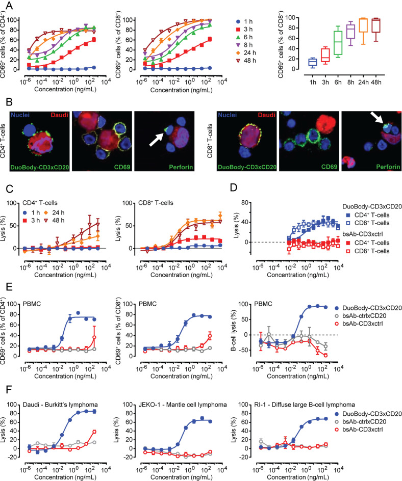
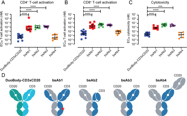
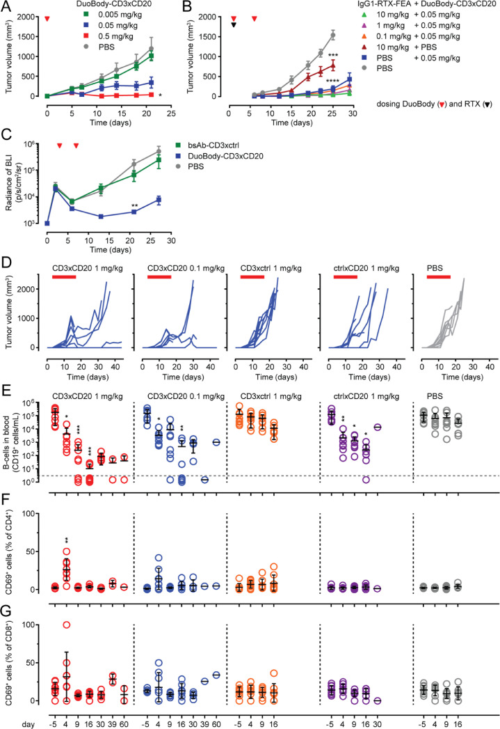
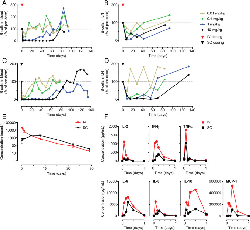
Findings: DuoBody-CD3xCD20 induced highly potent T-cell activation and T-cell-mediated cytotoxicity towards malignant B cells in vitro. Comparison of DuoBody-CD3xCD20 to CD3 bsAb targeting alternative B-cell antigens, or to CD3xCD20 bsAb generated using alternative CD20 Ab, emphasized its exceptional potency. In vitro comparison with other CD3xCD20 bsAb in clinical development showed that DuoBody-CD3xCD20 was significantly more potent than three other bsAb with single CD3 and CD20 binding regions and equally potent as a bsAb with a single CD3 and two CD20 binding regions. DuoBody-CD3xCD20 showed promising anti-tumour activity in vivo, also in the presence of excess levels of a CD20 Ab that competes for binding. In cynomolgus monkeys, DuoBody-CD3xCD20 demonstrated profound and long-lasting B-cell depletion from peripheral blood and lymphoid organs, which was comparable after subcutaneous and intravenous administration. Peak plasma levels of DuoBody-CD3xCD20 were lower and delayed after subcutaneous administration, which was associated with a reduction in plasma cytokine levels compared to intravenous administration, while bioavailability was comparable.

Interpretation: Based on these preclinical studies, a clinical trial was initiated to assess the clinical safety of subcutaneous DuoBody-CD3xCD20 in patients with B-cell malignancies.

Funding: Genmab.

Keywords: B cell malignancy; Bispecific antibody; CD20; CD3; Subcutaneous administration; T cell redirection.


Figures

Fig. 1
Fig. 1
Functional characterization of DuoBody-CD3xCD20 in vitro. A: Isolated T cells were incubated with DuoBody-CD3xCD20 and Daudi cells (E:T ratio 2:1). Percentages of CD69-positive CD4+ (left) and CD8+T cells (middle) were determined by flow cytometry at the indicated time points. Data for one representative donor out of four donors is shown. Graphs show the mean ± standard error of the mean (SEM) of duplicate wells. The right panel shows percentages of CD69-positive CD4+T cells. Data shown are pooled data from 6–8 donors for T cells treated with 3.3 ng/mL DuoBody-CD3xCD20. Boxes extend from 25th to 75th percentiles. Whiskers are drawn down to minimum and up to maximum percentages. Median percentages are indicated. A significant linear trend was observed in time (one-way ANOVA with post-test for linear trend). B: Isolated T cells were co-cultured with Daudi cells (E:T ratio 1:1) in the presence of 1 µg/mL DuoBody-CD3xCD20 for 24 h. Images were taken at a 60x magnification (plus extra optical magnification of the left pictures) and are representative of three experiments. C: Isolated CD4+ (left) or CD8+T cells (right) were incubated with DuoBody-CD3xCD20 and chromium-labelled Daudi cells (E:T ratio 20:1; this relatively high E:T ratio was chosen to make sure that a signal could be measured for CD4+T cells and at early time points) for the indicated time periods. Data shown are mean percentages of Daudi cell lysis ± SEM of triplicates. Data from one representative donor out of six donors tested is shown. D: Isolated CD4+ or CD8+T cells were incubated with DuoBody-CD3xCD20 or bsAb-CD3xctrl and chromium-labelled Daudi cells (E:T ratio 8:1) for 24 h. Data shown are mean percentages of Daudi cell lysis ± SEM of triplicates. Data from one representative experiment out of 3 performed is shown. E: PBMC were incubated with indicated Ab for 72 h. Percentages of CD69-positive CD4+ (left) and CD8+T cells (middle) and the remaining number of intact endogenous B cells (right) were determined by flow cytometry. Data shown are from one representative experiment out of four performed and are mean percentages ± SEM of duplicate wells. F: Isolated T cells were incubated with DuoBody-CD3xCD20, bsAb-CD3xctrl or bsAb-ctrlxCD20 and indicated B-cell lymphoma cell lines (E:T ratio 2:1) for 48 h. The remaining number of intact lymphoma cells (right) were determined by flow cytometry. Data shown for each cell line are from one representative donor (for number of donors tested see Table 1) and are mean percentages ± SEM of duplicate wells.
Fig. 2
Fig. 2
Assessment of the contribution of the B-cell-targeting Fab arm to CD3 bsAb efficacy in vitro. A: CD3 bsAb targeting eight different B-cell antigens were added at a concentration of 1 µg/mL to a coculture of purified T cells and chromium-loaded target cells (E:T ratio 10:1). Target cell lysis was measured by chromium release after 24 h. The B-cell antigens are depicted in order from lowest to highest expression level on the B-cell line used as target cell in the assay (CD70: 3,900 sABC on RPMI-8226; CD24: 4,000 sABC on RPMI-8226; CD22: 16,000 sABC on RI-1; CD79b: 111,000 sABC on Daudi; HLA-DR: 205,000 sABC on Daudi; CD37: 224,000 sABC on OCI-Ly7; CD20: 240,000 sABC on RI-1, CD138: 697,000 sABC on RPMI-8226; as determined by QiFi analysis). Experiments were performed twice in triplicates, with two different T-cell donors per experiment (except for CD3xCD37: one experiment with two donors; CD3xCD20: three experiments with two donors; CD3xCD38: two experiments with two donors and one experiment with one donor). Figure shows the mean ± SD for one donor from one representative experiment performed in triplicate. B: Binding of indicated CD3xCD20 (left), CD3xCD37 (middle) and CD3xCD70 (right) bsAb to their TAA on Daudi cells was measured by flow cytometry. Data is shown as mean fluorescence intensities (MFI). Graphs show one representative experiment out of 2 (CD3xCD37, CD3xCD70) or 3 (CD3xCD20) performed. C: Isolated T cells were incubated with indicated CD3 bsAb or control Ab (as indicated) and chromium-labelled Daudi cells (E:T ratio 10:1) for 24 h. T-cell-mediated cytotoxicity is shown as the mean percentage of Daudi cell lysis ± SEM of triplicates. One representative donor out of 4 donors tested in 2 separate experiments is shown.
Fig. 3
Fig. 3
In vitro comparison of DuoBody-CD3xCD20 with other CD3xCD20 bsAb in clinical development. A-C: Isolated T cells were incubated with Ab and CD20-expressing Daudi cells (E:T ratio 2:1). Percentages CD69-positive CD4+ (A) and CD8+ (B) cells and cytotoxic activity (C) were assessed by flow cytometry after 48 h. Each dot represents the EC50 value calculated from one experiment. Different T-cell donors were used for the individual experiments. Median EC50 values ± interquartile range for each antibody are indicated. D. Cartoon of DuoBody-CD3xCD20 and bsAb1-4. DuoBody-CD3xCD20,obtained by cFAE, is an IgG1 and contains the L234F, L235E, D265A Fc-silencing mutations; bsAb1, obtained by asymmetric reengineering technology, has a common light chain, is an IgG4 with S228P hinge stabilization and E233P-F234V-L235A-G236del Fc-silencing mutations and mutations in one of the Fc regions to purify bsAb (H435R, Y436F; indicated by star); bsAb2 is an XmAb (Fab-FcxFv-Fc) IgG1 with E233P-L234V-L235A-G236del-S267K Fc-silencing mutations, bsAb3 is a knob-into-hole IgG1 with an N297G Fc-silencing mutation, bsAb4 is a Fab-Fab-FcxFab-Fc (2:1) knob-into-hole CrossMab IgG1 with L234A-L235A-P329G Fc-silencing mutations (references for sequences of these bsAb are mentioned in the Mat & Meth section; formats are reviewed in [46]).
Fig. 4
Fig. 4
Anti-tumour activity of DuoBody-CD3xCD20 in vivo in humanized mouse models. A-B: Tumours were induced in NOD-SCID mice by SC injection of a 1:1 mixture of human PBMC and Raji-luc cells at day 0. Immediately after tumour inoculation, mice (5 per group) were treated with a single IV dose of DuoBody-CD3xCD20 (0.5, 0.05, 0.005 mg/kg) (A) or mice (10 per group) were given a single IV dose of IgG1-RTX-FEA (10, 1 or 0.1 mg/kg), followed by IV treatment with 0.05 mg/kg DuoBody-CD3xCD20 4 h later, treatment with DuoBody-CD3xCD20 was repeated on day 7 (B). Data shown are mean tumour volumes per treatment group ± SEM. Differences were analysed by Mann-Whitney two-tailed test versus PBS (A: day 21; B: day 25). Statistically significant differences are indicated as follows: * p < 0.05, *** p < 0.0005, **** p < 0.0001 (A-B). C: Tumours were induced in BRGS-HIS mice by IV injection of Daudi-luc cells at day 0. Mice were treated IV with 1 mg/kg DuoBody-CD3xCD20 on day 3 and day 7. Tumour growth was evaluated by BLI and depicted as average radiance of BLI ± SEM (7 mice per group). Statistically significant differences compared to PBS-treated mice were determined by Kruskal-Wallis ANOVA test followed by Dunn's multiple comparison post-test (** p < 0.01). D-G: Tumours were induced in BRGS-HIS mice by SC injection of Raji-luc cells at day 0. Mice were treated IV with 1 mg/kg or 0.1 mg/kg bsAb or vehicle control on days 3, 7, 10, 14, and 17. Tumour growth is shown for each individual mouse, one graph per treatment group (D). Individual (circles) and average (black line) absolute numbers of circulating human CD19+B cells determined by flow cytometry at indicated time points (E). T-cell activation was determined on indicated time points by flow cytometry. Individual (circles) and average (black line) percentages of CD69+ cells in CD4+T-cell population (F) and CD8+T-cell population (G) are shown. Statistically significant differences compared to PBS-treated mice at each time point were determined by Kruskal-Wallis ANOVA test with Dunn's multiple comparisons test (*p < 0.05, ** p < 0.01, *** p < 0.001) (E-G).
Fig. 5
Fig. 5
Anti-B-cell activity of DuoBody-CD3xCD20 in cynomolgus monkeys after IV and SC administration. A-D: B-cell counts (CD19+ cells) are depicted per monkey (n = 2 per dose group) as a percentage of the B-cell counts prior to dosing. B-cell counts in peripheral blood (A, C) and lymph nodes (B, D) were measured by flow cytometry and are shown for the indicated dose levels of a single IV (A, B) or SC (C, D) administration of DuoBody-CD3xCD20. E: Mean ± SEM plasma concentration profiles for DuoBody-CD3xCD20 measured by Single Molecule Counting ImmunoAssay (SMCIA) after a single IV or SC dose of 1 mg/kg DuoBody-CD3xCD20 (6 monkeys/group). F: Mean plasma concentration profiles of indicated cytokines measured after a single IV or SC dose of 1 mg/kg DuoBody-CD3xCD20 (6 monkeys/group).

References

    1. Guerra V.A., Jabbour E.J., Ravandi F., Kantarjian H., Short N.J. Novel monoclonal antibody-based treatment strategies in adults with acute lymphoblastic leukemia. Ther Adv Hematol. 2019;10 - PMC - PubMed
    1. Huguet F., Tavitian S. Emerging biological therapies to treat acute lymphoblastic leukemia. Expert Opin Emerg Drugs. 2017;22:107–121. - PubMed
    1. Owen C.J., Stewart D.A. Obinutuzumab for B-cell malignancies. Expert Opin Biol Ther. 2014;14:1197–1205. - PubMed
    1. Gisselbrecht C., Glass B., Mounier N. Salvage regimens with autologous transplantation for relapsed large B-cell lymphoma in the rituximab era. J Clin Oncol. 2010;28:4184–4190. - PMC - PubMed
    1. Johnson N.A., Leach S., Woolcock B. CD20 mutations involving the rituximab epitope are rare in diffuse large B-cell lymphomas and are not a significant cause of R-Chop failure. Haematologica. 2009;94:423–427. - PMC - PubMed

MeSH terms

LinkOut - more resources