. 2020 Aug;146(2):377-389.e10.
doi: 10.1016/j.jaci.2020.01.013. Epub 2020 Jan 23.

Charcot-Leyden crystal protein/galectin-10 interacts with cationic ribonucleases and is required for eosinophil granulogenesis

Affiliations

Charcot-Leyden crystal protein/galectin-10 interacts with cationic ribonucleases and is required for eosinophil granulogenesis

Milica M Grozdanovic et al. J Allergy Clin Immunol. 2020 Aug.

Abstract

Background: The human eosinophil Charcot-Leyden crystal (CLC) protein is a member of the Galectin superfamily and is also known as galectin-10 (Gal-10). CLC/Gal-10 forms the distinctive hexagonal bipyramidal crystals that are considered hallmarks of eosinophil participation in allergic responses and related inflammatory reactions; however, the glycan-containing ligands of CLC/Gal-10, its cellular function(s), and its role(s) in allergic diseases are unknown.

Objective: We sought to determine the binding partners of CLC/Gal-10 and elucidate its role in eosinophil biology.

Methods: Intracellular binding partners were determined by ligand blotting with CLC/Gal-10, followed by coimmunoprecipitation and coaffinity purifications. The role of CLC/Gal-10 in eosinophil function was determined by using enzyme activity assays, confocal microscopy, and short hairpin RNA knockout of CLC/Gal-10 expression in human CD34+ cord blood hematopoietic progenitors differentiated to eosinophils.

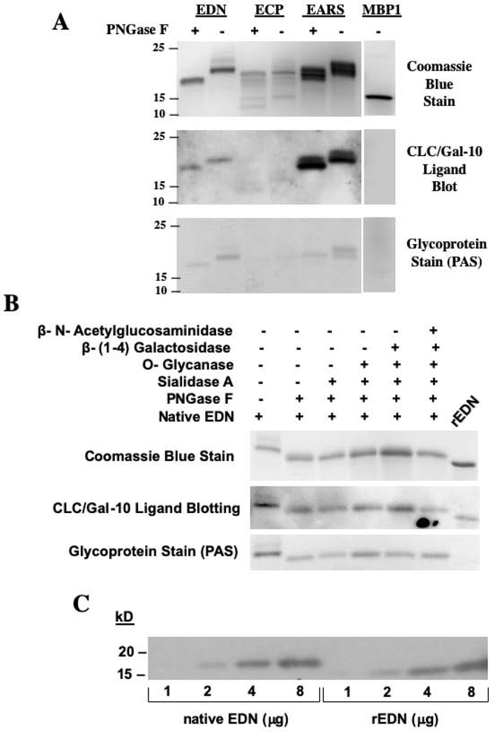
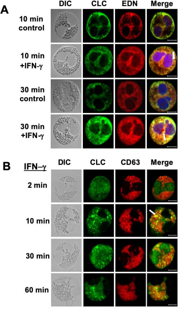
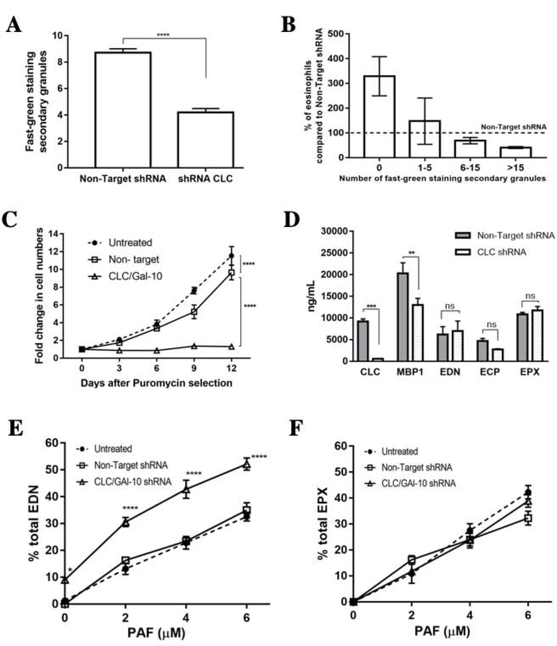
Results: CLC/Gal-10 interacts with both human eosinophil granule cationic ribonucleases (RNases), namely, eosinophil-derived neurotoxin (RNS2) and eosinophil cationic protein (RNS3), and with murine eosinophil-associated RNases. The interaction is independent of glycosylation and is not inhibitory toward endoRNase activity. Activation of eosinophils with INF-γ induces the rapid colocalization of CLC/Gal-10 with eosinophil-derived neurotoxin/RNS2 and CD63. Short hairpin RNA knockdown of CLC/Gal-10 in human cord blood-derived CD34+ progenitor cells impairs eosinophil granulogenesis.

Conclusions: CLC/Gal-10 functions as a carrier for the sequestration and vesicular transport of the potent eosinophil granule cationic RNases during both differentiation and degranulation, enabling their intracellular packaging and extracellular functions in allergic inflammation.

Keywords: Charcot-Leyden; ECP; EDN; Eosinophils; RNase 2; RNase 3; galectins; granulogenesis; ribonucleases.


Conflict of interest statement

Conflict of Interest

The authors have no financial or other conflicts of interest to declare.

Figures

Figure 1.
Figure 1.. CLC/Gal-10 interacts with eosinophil granule cationic ribonucleases.
(A, B) CLC/Gal-10 was used as a probe to “ligand blot” blood eosinophil lysate, followed by detection with anti-CLC/Gal-10 antibody. CLC/Gal-10 binds to an ~21kD band in blood eosinophil lysate that is reduced to ~18 kD upon digestion of the lysates with PNGase F. (C) EDN co-purifies with CLC/Gal-10 from lysates of AML14.3D10 eosinophils or purified blood eosinophils. Samples affinity purified over an anti-CLC/Gal-10 antibody column were blotted with either anti-CLC/Gal-10 or anti-EDN/ECP (cross-reactive) antibodies. Purified MBP-1 and anti-MBP-1 antibodies were used as a control. (D) EDN is co-immunoprecipitated by anti-CLC/Gal-l0 antibody (left panel) and CLC/Gal-10 is co-immunoprecipitated by anti-EDN antibody (right panel) from AML14.3D10 eosinophil lysate. The initial AML14.3D10 lysate (input) was included as a positive control. The immunoprecipitations were performed using rabbit non-immune (NI) IgG, anti-CLC/Gal-10, or anti-EDN antibodies.
Figure 2.
Figure 2.. Interaction of CLC/Gal-10 with the cationic endoribonucleases is not glycan-dependent.
(A) Purified human (EDN, ECP, MBP-1) and murine (EARS) eosinophil granule proteins (2.5 μg/ each) bind CLC/Gal-10 in ligand blot, with or without prior PNGase digestion. Electrotransferred samples were detected by Coomassie Blue stain (top), anti-CLC/Gal-10 antibody (middle) or glycoprotein stain (PAS) (bottom). (B) Native (glycosylated) human EDN binds CLC/Gal-10 even after being subjected to sequential digestions with PNGase F, Sialidase A, O-Glycanase, β-(1–4) Galactosidase, and β-N-Acetylglucosaminidase. The samples were stained for protein by Coomassie Blue (top), ligand blotted using CLC/Gal-10 followed by anti-CLC/Gal-10 (middle), and glycoprotein by PAS (bottom). (C) Purified native EDN and recombinant EDN have similar affinities for CLC/Gal-10 binding, as demonstrated by ligand blotting increasing amounts of EDN (1, 2, 4, and 8 μg) with crystal-derived CLC/Gal-10, followed by detection of bound CLC/Gal-10 with anti-CLC/Gal-10 antibodies. Bacterially expressed non-glycosylated rEDN was included for comparison.
Figure 3.
Figure 3.. CLC/Gal-10 does not inhibit the ribonuclease activity of EDN.
(A) 80 pg of purified native EDN was analyzed for RNase activity in the absence or presence of increasing amounts of CLC/Gal-10 protein, or placental RNase inhibitor. The relative fluorescence units reflect the amount of fluorescence emitted by the cleavable fluorescent-labeled RNase substrate. The amount of crystal-derived CLC/Gal-10 protein ranged from 0 to 1600 pg, and human placental RNase inhibitor ranged from 0 to 5 units. (B) Increasing amounts of EDN were incubated with a constant amount of either CLC/Gal-10 (400 pg) or placental RNase inhibitor (5 units). Results are representative of 3 independent experiments with three different preparations of CLC/Gal-10 protein purified by crystallization from blood eosinophils. ns = not significant
Figure 4.
Figure 4.. Activation of blood eosinophils with IFN-γ induces the intracellular co-localization of CLC/Gal-10 with EDN and CD63.
(A) Representative confocal images of blood eosinophils cultured without (control) or with IFN-γ (500 U/ml) for periods of 10 to 30 min. Upon activation with IFN-γ, the merged images clearly display yellow regions indicative of co-localization of CLC/Gal-10 and EDN. Co-localization reaches maximum levels 30 min after activation and dissipates after 60 min (not shown). (B) Purified eosinophils stimulated with IFN-γ (500 U/ml) for periods of 2, 10, 30, or 60 minutes show CLC/Gal-10 and CD63 co-localization at discrete punctate sites in the cytosol consistent in size with eosinophil secondary granules. Maximum colocalization is visible 30 min after activation. DIC shows the appearance of typical eosinophil secondary granules in the cytosol. Results include representative images from 4 independent experiments. Arrows highlight pockets of co-localization. White size bars in lower right corner indicate 5μm.
Figure 5.
Figure 5.. shRNA knock-down of CLC/Gal-10 in cord-blood derived eosinophil progenitor cells leads to impaired eosinophil differentiation/ granulogenesis.
(A) Purified human CD34+ cord blood-derived progenitor cells were transduced with specific CLC/Gal-10 shRNA or Non-Target control shRNA and then differentiated toward the eosinophil lineage with IL-5. Immunofluorescence staining of cells at day 14 shows an almost complete loss of CLC/Gal-10 expression in cells treated with CLC/Gal-10 specific shRNA (Fig. 5A, top panel). Fast Green/Neutral Red staining of cells shows characteristic features of mature eosinophils, including red nuclei, pink cytoplasm and turquoise green granules, confirming that the cells were successfully differentiated toward the eosinophil lineage (middle panel). After 14 days, the CLC/Gal-10 knock-down cells display a significant reduction in the number of Fast Green stained secondary granules (middle panel), and cells stained on day 21 show increasing differences in cellular morphology as compared to control cells (bottom panel), with large empty granules (arrow). (B) Representative images of Fast Green/ Neutral Red stained cells 21 days post transduction. Cells transduced with CLC/Gal-10 specific shRNA manifest predominantly with large, empty granules. Images are representative of 2 independent experiments performed in triplicate. Size bars represent 5 μm.
Figure 6.
Figure 6.. CLC/Gal-10 deficient (knock-down) eosinophils have fewer secondary granules, a non-proliferative phenotype, decreased MBP-1 expression, and increased secretion of EDN in response to PAF stimulation.
(A) CLC/Gal-10 knock-down cells display an ~42% decrease in average number of secondary granules as compared to non-target shRNA transduced cells. (B) Cell populations transduced with CLC/Gal-10 shRNA show a dramatic increase of cells with no detectable granules and a strikingly smaller number of cells with more than 15 granules. (C) CLC-deficient cells display a non-proliferative phenotype, in contrast to non-target shRNA treated cells that continued to proliferate throughout the 21 days, although at a slower pace than untreated cells (expected being under Puromycin selection). (D) CLC/Gal-10 knock-down causes a significant decrease in ELISA detected levels of CLC/Gal-10 and MBP-1 in cell lysates, but no significant change in EDN, ECP, and EPX expression. CLC-deficient eosinophils still exhibit dose-dependent secretion of EDN (E) and EPX (F) when activated with the secretagogue PAF. The secretion of EPX (F) by CLC-deficient eosinophils was not significantly different from untreated or non-target shRNA control cells; however, amounts of secreted EDN (E) was significantly higher in CLC-deficient eosinophils. Results represent mean (200 counted cells per treatment group) ± SEM from 2 independent experiments. (ns = not significant,**p ≤ 0.01, ***p ≤ 0.001, ****p ≤ 0.0001).

References

    1. Charcot JM, Robin C. Observation de Leukocythemia. C R Mem Soc Biol 1853; 5:44.
    1. von Leyden EV. Zur Kenntniss des Bronchial Asthma. [Virchows] Archiv für pathologische Anatomie und Physiologie, und für klinische Medizin 1872; 54:324–44.
    1. Ackerman SJ, Zhou Z-Q, Tenen DG, Clark MA, Tu Y-P, Irvin CG. Human eosinophil lysophospholipase (Charcot-Leyden crystal protein): Molecular cloning, expression, and potential functions in asthma In: Gleich GJ, Kay AB, editors. Eosinophils In Allergy and Inflammation. New York: Marcel Dekker; 1994. p. 21–54.
    1. Ackerman SJ. Characterization and functions of eosinophil granule proteins In: Makino S, Fukuda T, editors. Eosinophils: Biological and Clinical Aspects. Boca Raton: CRC Press; 1993. p. 33–74.
    1. Gleich GJ, Loegering DA, Mann KG, Maldonado JE. Comparative properties of the Charcot-Leyden crystal protein and the major basic protein from human eosinophils. J Clin Invest 1976; 57:633–40. - PMC - PubMed

Publication types