. 2019 Jul:116:282-291.
doi: 10.1016/j.trac.2019.04.023. Epub 2019 Apr 30.

Computational Insights into Compaction of Gas-Phase Protein and Protein Complex Ions in Native Ion Mobility-Mass Spectrometry

Affiliations

Computational Insights into Compaction of Gas-Phase Protein and Protein Complex Ions in Native Ion Mobility-Mass Spectrometry

Amber D Rolland et al. Trends Analyt Chem. 2019 Jul.

Abstract

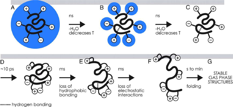
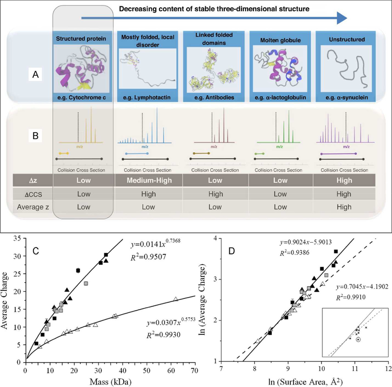
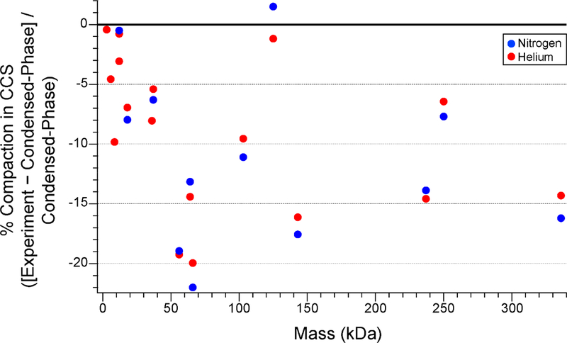
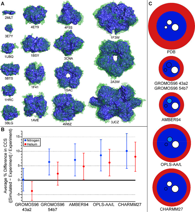
Native ion mobility-mass spectrometry (IM-MS) is a rapidly growing field for studying the composition and structure of biomolecules and biomolecular complexes using gas-phase methods. Typically, ions are formed in native IM-MS using gentle nanoelectrospray ionization conditions, which in many cases can preserve condensed-phase stoichiometry. Although much evidence shows that large-scale condensed-phase structure, such as quaternary structure and topology, can also be preserved, it is less clear to what extent smaller-scale structure is preserved in native IM-MS. This review surveys computational and experimental efforts aimed at characterizing compaction and structural rearrangements of protein and protein complex ions upon transfer to the gas phase. A brief summary of gas-phase compaction results from molecular dynamics simulations using multiple common force fields and a wide variety of protein ions is presented and compared to literature IM-MS data.

Keywords: electrospray ionization; gas phase; ion structure; molecular dynamics; native ion mobility-mass spectrometry; protein.


Conflict of interest statement

Competing Interests Statement. The authors declare no competing interests.

Figures

Figure 1.
Figure 1.
(A-D) Depiction of charging and self-solvation of charge sites (+ and − symbols) for globular protein ions during evaporation of the nanoelectrospray droplet on the nanosecond to picosecond timescale and (E-G) subsequent structural rearrangement and unfolding at longer timescales. Figure copyright 2008 National Academy of the Sciences, reproduced with permission from the National Academy of the Sciences.
Figure 2.
Figure 2.
(A) Schematic illustration of condensed-phase protein structure types and (B) typical charge state and CCS distributions in IM-MS experiments. (C) Average charge states for protein ions formed under non-denaturing (native) conditions (open triangles) and denaturing conditions (all other symbols) as a function of mass and (D) relationships between average charge state and condensed-phase surface area for the same ions. A, B reprinted from Current Opinion in Chemical Biology, v. 42, D. Stuchfield and P. Barran, “Unique insights into intrinsically disordered proteins provided by ion mobility mass spectrometry,” pp. 177–185, copyright 2018, with permission from Elsevier. C, D reprinted with permission from Analytical Chemistry, v. 83, L. Testa, S. Brocca, and R. Grandori, “Charge-Surface Correlation in Electrospray Ionization of Folded and Unfolded Proteins,” pp. 6459–6463, copyright 2011 American Chemical Society.
Figure 3.
Figure 3.
Plot of fractional compaction of protein and protein complex ions produced by nESI under native conditions measured by IM-MS in He or N2 buffer gas as compared to CCSs for condensed-phase structures computed using Collidoscope [86]. Experimental CCSs from ref. [84] and [85]. Protein Data Bank identifiers for all protein structures are listed in Figure 4.
Figure 4.
Figure 4.
(A) Structures of protein and protein complex ions from Fig. 3 before (green mesh) and after (blue solid) MD simulation of gas-phase compaction using GROMOS96 43a2 FF (see text). Protonated ions simulated (with Protein Data Bank identifiers indicated below structures) are melittin4+ (2MLT), insulin3+ (3E7Y), ubiquitin5+ (1UBQ), insulin dimer5+ (5BTS), cytochrome c7+ (1HRC), β-lactoglobulin monomer7+ (3BLG), insulin hexamer10+ (4EY9), transthyretin tetramer15+ (1F41), avidin tetramer16+ (1AVE), bovine serum albumin15+ (4F5S), concanavalin A tetramer21+ (3CNA), serum amyloid P component pentamer24+ (1SAC), alcohol dehydrogenase tetramer24+ (4W6Z), pyruvate kinase tetramer32+ (1F3W), serum amyloid P component decamer33+ (2A3W), glutamate dehydrogenase hexamer40+ (3JCZ). (B) Plot of average percent difference between experimental CCS data from ref. [84] and [85] and CCSs computed for MD-compacted ions shown in A using Collidoscope for each of the 5 FFs tested (see text). (C) Schematic depiction of typical degree of surface (red) and interior (dark blue) compaction predicted by MD simulations using 5 different FFs for ions represented in A and B. Embedded circles represent the typical size (small: 5–12 Å diameter, medium: 12–25 Å, and large: ≥ 25 Å) of cavities in the ions that are fully eliminated (dark blue), sometimes eliminated (light blue), or not eliminated (white) during the MD simulations.

References

    1. Heck AJR, van den Heuvel RHH, Investigation of Intact Protein Complexes by Mass Spectrometry, Mass Spectrom. Rev 23 (2004) 368–389. - PubMed
    1. Konijnenberg A, Butterer A, Sobott F, Native Ion Mobility-Mass Spectrometry and Related Methods in Structural Biology, Biochim. Biophys. Acta, Proteins Proteomics 1834 (2013) 1239–1256. - PubMed
    1. Gabelica V, Marklund E, Fundamentals of Ion Mobility Spectrometry, Curr.Opin. Chem. Biol 42 (2018) 51–59. - PubMed
    1. Testa L, Brocca S, Grandori R, Charge-Surface Correlation in Electrospray Ionization of Folded and Unfolded Proteins, Anal. Chem 83 (2011) 6459–6463. - PubMed
    1. Benesch JLP, Ruotolo BT, Simmons DA, Robinson CV, Protein Complexes in the Gas Phase: Technology for Structural Genomics and Proteomics, Chem. Rev 107 (2007) 3544–3567. - PubMed

LinkOut - more resources