Review
. 2020 Mar;294(1):177-187.
doi: 10.1111/imr.12838. Epub 2020 Jan 27.

Immunometabolism in the development of rheumatoid arthritis

Affiliations
Review

Immunometabolism in the development of rheumatoid arthritis

Cornelia M Weyand et al. Immunol Rev. 2020 Mar.

Abstract

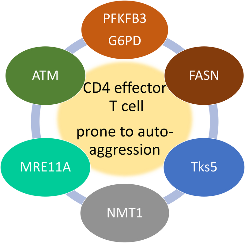
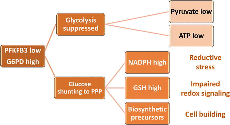
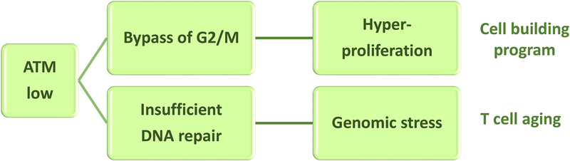
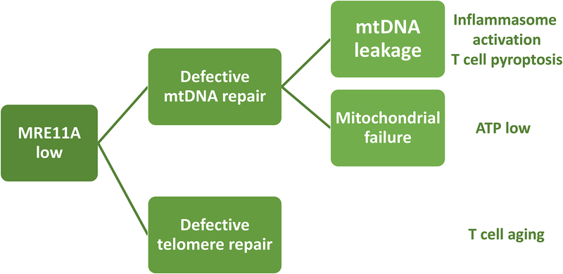
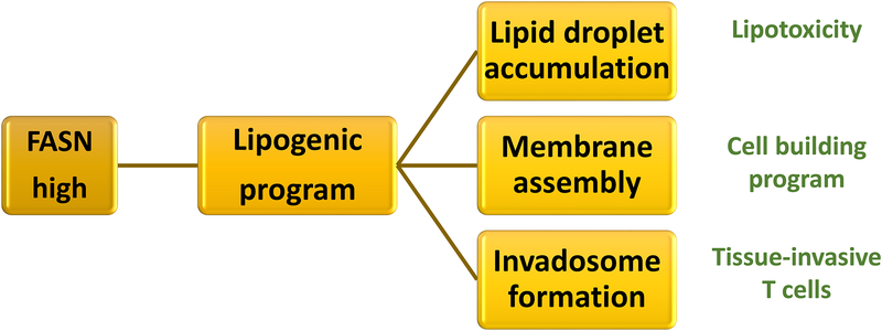
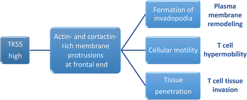
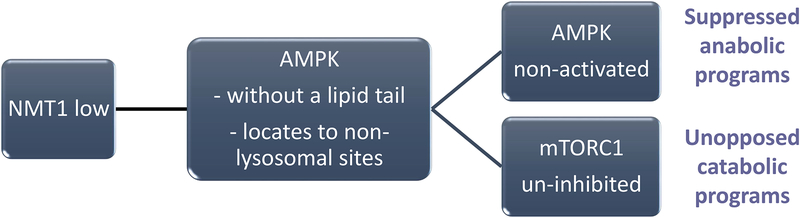
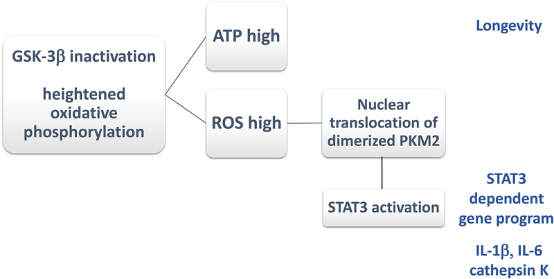
In rheumatoid arthritis (RA), breakdown of self-tolerance and onset of clinical disease are separated in time and space, supporting a multi-hit model in which emergence of autoreactive T cells is a pinnacle pathogenic event. Determining factors in T cell differentiation and survival include antigen recognition, but also the metabolic machinery that provides energy and biosynthetic molecules for cell building. Studies in patients with RA have yielded a disease-specific metabolic signature, which enables naive CD4 T cells to differentiate into pro-inflammatory helper T cells that are prone to invade into tissue and elicit inflammation through immunogenic cell death. A typifying property of RA CD4 T cells is the shunting of glucose away from glycolytic breakdown and mitochondrial processing toward the pentose phosphate pathway, favoring anabolic over catabolic reactions. Key defects have been localized to the mitochondria and the lysosome; including instability of mitochondrial DNA due to the lack of the DNA repair nuclease MRE11A and inefficient lysosomal tethering of AMPK due to deficiency of N-myristoyltransferase 1 (NMT1). The molecular taxonomy of the metabolically reprogrammed RA T cells includes glycolytic enzymes (glucose-6-phosphate dehydrogenase, phosphofructokinase), DNA repair molecules (MRE11A, ATM), regulators of protein trafficking (NMT1), and the membrane adapter protein TSK5. As the mechanisms determining abnormal T cell behavior in RA are unraveled, opportunities will emerge to interject autoimmune T cells by targeting their metabolic checkpoints.

Keywords: DNA damage; DNA repair; T cell; autoimmunity; cell cycle; glycolysis; macrophage; mitochondria; myristoylation; protein trafficking; rheumatoid arthritis; telomere.


Figures

Figure 1.
Figure 1.. Metabolic checkpoints in pro-inflammatory and auto-aggressive T cells.
Studies in CD4 T cells from patients with RA have identified a series of molecules that deviate T cell function towards pro-inflammatory capabilities. All molecules identified have in common that they regulate or are regulated by the cell’s metabolic machinery. PFKFB3, G6PD and FASN directly regulate cytosolic glycolysis and lipogenesis. The cell cycle kinase ATM senses metabolic activity through reactive oxygen species to coordinate cell cycle passage to nutrient supply. The DNA repair nuclease MRE11A maintains metabolic competence by protecting mitochondrial DNA. The transferase NMT1 enables trafficking of the energy sensor AMPK to the lysosomal surface. Metabolic intermediates regulate expression of the membrane adaptor molecule Tks5, thereby rendering T cells tissue-invasive.
Figure 2.
Figure 2.. Glucose shunting to the pentose phosphate pathway enables anabolic processes.
In RA CD4 T cells, glucose is shunted from glycolytic breakdown towards the pentose phosphate pathway (PPP). Transcriptional repression of phosphofructokinase/fructose biphosphatase 3 (PFKFB3) results in reduced ATP and pyruvate production. Upregulation of Glucose-6-phosphate dehydrogenase (G6PD) funnels glucose towards the PPP, supplying NADPH, reduced glutathione (GSH) and biosynthetic precursors. As an outcome, the cell’s redox status shifts towards reductive stress, impairing redox-dependent signaling. Also, the cell has access to biosynthetic precursors, enabling a cell building program.
Figure 3.
Figure 3.. Dysfunctional cell cycle control results in T cell hyperproliferation.
In RA CD4 T cells, the DNA damage sensing kinase Ataxia telangiectasia mutated (ATM) is impaired. ATM is required for the initiation of double-strand break repair by homologous recombination and functions by slowing down the G2/M phase of the cell cycle. In patient-derived CD4 T cells, ATM protein and activity are diminished. ATM function requires dimerization, which is dependent on redox signaling. Low abundance of reactive oxygen species in RA T cells results in insufficient ATM activation. The outcome includes a high DNA damage burden, bypassing of the G2/M cell cycle checkpoint and T cell hyperproliferation.
Figure 4.
Figure 4.. DNA repair failure in the mitochondria and at the telomere causes metabolic maladaptation and premature T cell aging.
In RA CD4 T cells, the DNA repair is inefficient, affecting both the mitochondrial genome and the telomeric ends. The nuclease MRE11A is a limiting factor in double strand break repair and is transcriptionally repressed in RA CD4 T cells. Lack of MRE11A’s nucleolytic activity in the mitochondria leads to instability of mitochondrial DNA (mtDNA), leakage of mtDNA into the cytosol, activation of the inflammasome and induction of immunogenic T cell death (pyroptosis). Lack of mitochondrial MRE11A leads to diminished oxygen consumption and ATP generation. Insufficiency of MRE11A at the telomeric ends results in telomere fragility and T cell aging.
Figure 5.
Figure 5.. Disproportional lipogenesis fuels tissue invasiveness of RA T cells.
RA CD4 T cells favor lipogenesis over lipolysis. The cytosol is rich in reducing equivalents (NADPH), mitochondrial b-oxidation is impaired and the lipogenic gene signature, e.g. fatty acid synthase (FASN) is upregulated; all supporting the synthesis and not the breakdown of fatty acids. Excess lipogenesis leads to the deposition of cytosolic lipid droplets, availability of lipid precursors to build membranes and the formation of invasive membranes structures resembling invadosomes. As a result, T cells enter a cell building program and become tissue-invasive.
Figure 6.
Figure 6.. Membrane ruffling enables T cell tissue invasiveness.
CD4 T cells from RA patients have high abundance of Tks5 (encoded by the SH3PXD2A gene), a SH3- and PX-domain containing scaffolding protein. Tks5 localizes to the podosmal membrane extrusions on the frontal end of T cells. Formation of membrane ruffles is part of a metabolically-controlled T cell motility program, and high expression of Tks5 renders T cells highly mobile, permitting transition from blood vessels into the extravascular space and fast maneuvering in extracellular matrix.
Figure 7.
Figure 7.. Compromised myristoylation alters subcellular trafficking of AMP-activated protein kinase (AMPK).
In RA T cells, posttranslational lipidation modification of proteins is compromised due to impaired function of the N-myristoyltransferase 1 (NMT1). Lack of NMT1 function affects the subcellular distribution of the energy sensor AMPK, which is normally recruited to the cytosolic face of the lysosome to monitor the AMP/ATP ratio. Energy deficiency prompts AMPK activation, triggers a multitude of anabolic programs and inhibits energy-utilizing catabolic processes by inactivating mTORC1. In RA CD4 T cells, AMPK lacks a lipid tail, fails to relocate to the lysosomal surface and malfunctions as an mTORC1 inhibitor. AMPK deficiency promotes mitochondrial restraint and confers continuous mTORC1 activity.
Figure 8.
Figure 8.. Hypermetabolic macrophages in rheumatoid arthritis.
In RA macrophages, Glycogen Synthase Kinase 3β (GSK-3β) is inactivated, enhancing the activity of the mitochondrial electron transport chain. GSK-3β is a constitutively active protein kinase that negatively regulates glucose homeostasis. Inactivation of GSK3β results in the activation of oxidative phosphorylation, enhanced ATP production and increased ROS release. Functional consequences include longevity of highly activated macrophages. ROS facilitate the dimerization of the cytosolic enzyme pyruvate kinase M2 (PKM2) and nuclear translocation of the enzyme, where it activates STAT3. Several pro-inflammatory activities of macrophages are dependent on GSK-3β inactivation and enhanced ROS release; such as the production of cathepsin K, IL-1β, and IL-6.

References

    1. Martins P, Fonseca JE. How to investigate: Pre-clinical rheumatoid arthritis. Best Pract Res Clin Rheumatol. 2019;101438. - PubMed
    1. Kalinkovich A, Gabdulina G, Livshits G. Autoimmunity, inflammation, and dysbiosis mutually govern the transition from the preclinical to the clinical stage of rheumatoid arthritis. Immunol Res. 2018;66(6):696–709. - PubMed
    1. Ma WT, Chang C, Gershwin ME, Lian ZX. Development of autoantibodies precedes clinical manifestations of autoimmune diseases: A comprehensive review. J Autoimmun. 2017;8395–112. - PubMed
    1. Deane KD, Demoruelle MK, Kelmenson LB, Kuhn KA, Norris JM, Holers VM. Genetic and environmental risk factors for rheumatoid arthritis. Best Pract Res Clin Rheumatol. 2017;31(1):3–18. - PMC - PubMed
    1. Nagy G, van Vollenhoven RF. Sustained biologic-free and drug-free remission in rheumatoid arthritis, where are we now? Arthritis Res Ther. 2015;17181. - PMC - PubMed

Publication types