. 2020 Mar;45(3):886-896.
doi: 10.3892/ijmm.2020.4474. Epub 2020 Jan 23.

MicroRNA‑137 suppresses the proliferation, migration and invasion of cholangiocarcinoma cells by targeting WNT2B

Affiliations

MicroRNA‑137 suppresses the proliferation, migration and invasion of cholangiocarcinoma cells by targeting WNT2B

Tengxiang Chen et al. Int J Mol Med. 2020 Mar.

Abstract

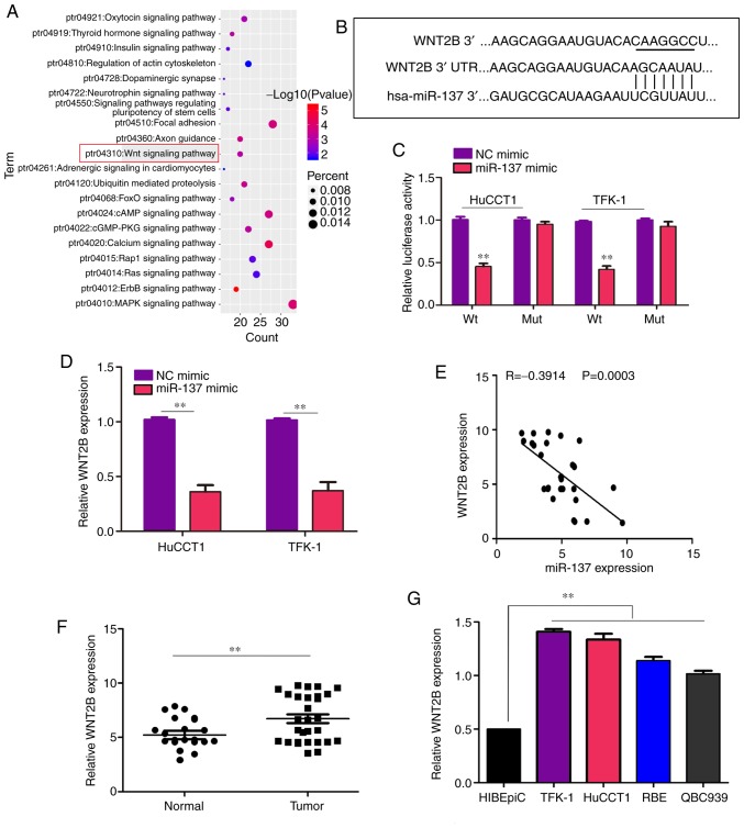
It is widely known that abnormal regulation of microRNAs (miRNAs/miRs) may contribute to the occurrence or development of tumors. The objective of the present study was to elucidate the function and underlying mechanism of miR‑137 in the progression of cholangiocarcinoma (CCA). The expression levels of miR‑137 in CCA tissues and cell lines were measured using reverse transcription‑quantitative PCR. The role of miR‑137 in the proliferation of CCA cells was assessed using the Cell Counting Kit‑8 assay, colony formation assay and cell cycle distribution analysis, while its effects on the migration and invasion of CCA cells were evaluated using Transwell assays. The function of miR‑137 on CCA growth in vivo was also investigated using a xenograft mouse model. Furthermore, the association between miR‑137 and Wnt family member 2B (WNT2B) was analyzed using bioinformatics, double luciferase assay and western blotting. It was verified that the expression of miR‑137 was low in CCA tissues and cell lines, whereas increased expression of miR‑137 significantly suppressed cell proliferation, decreased colony formation ability and induced G1 phase arrest. miR‑137 overexpression suppressed the migration and invasion ability of TFK‑1 and HuCCT1 cells. Furthermore, the results of the xenograft mouse model assays revealed that miR‑137 overexpression decreased tumor growth in vivo. The results of bioinformatics analysis and dual luciferase reporter assays demonstrated that WNT2B is directly regulated by miR‑137. The expression of WNT2B and Wnt‑pathway‑related proteins was decreased when miR‑137 was overexpressed. Restoring the expression of WNT2B notably reversed the inhibitory effect of miR‑137 on CCA cells. Therefore, the findings of the present study demonstrated that miR‑137 acts as a suppressor in CCA and inhibits CCA cell proliferation, migration and invasion through suppressing the expression of WNT2B.


Figures

Figure 1
Figure 1
miR-137 is significantly downregulated in cholangiocarcinoma. (A) The expression of miR-137 was detected in 29 cholangiocarcinoma tissues and 20 adjacent non-tumor tissues using reverse transcription-quantitative PCR. (B) The expression of miR-137 in the cholangiocarcinoma cell lines TFK-1, HuCCT1, RBE and QBC939 and HIBEpiCs was detected using reverse transcription-quantitative PCR. **P<0.01. miR, microRNA; HIBEpiCs, human intrahepatic biliary epithelial cells.
Figure 2
Figure 2
miR-137 represses cholangiocarcinoma cell proliferation in vitro. (A) Fluorescence microscope examination (magnification, ×200) and reverse transcription-quantitative PCR were used to detect the infection efficiency of miR-137 overexpression (LV-miR-137) and NC lentiviruses. (B) The effect of miR-137 on cholangiocarcinoma cell proliferation was detected by the Cell Counting Kit-8 assay. (C) The effect of miR-137 on cholangiocarcinoma cell colony formation ability was detected using colony formation assays. (D) Cell cycle distribution was analyzed after miR-137 overexpression in TFK-1 and HuCCT1 cells. (E) Western blotting was used to detect the expression of CDK2 and cyclin D1 in the miR-137 overexpression and normal control groups. GAPDH was used as the loading control. *P<0.05, **P<0.01. miR, microRNA; NC, negative control; LV, lentivirus; CDK, cyclin dependent kinase; OD, optical density.
Figure 3
Figure 3
miR-137 represses cholangiocarcinoma cell migration and invasion in vitro. (A) The effect of miR-137 on cholangiocarcinoma cell invasion and migration was examined by Transwell assays. Magnification, ×40. (B) Western blotting was used to detect the expression of N-cadherin and vimentin in the miR-137 overexpression and normal control groups. GAPDH was used as the loading control. **P<0.01. miR, microRNA; NC, negative control; LV, lentivirus; N, neural.
Figure 4
Figure 4
miR-137 inhibits tumor growth in vivo. (A) Representative images of subcutaneous tumors of the miR-137 overexpression and control groups. (B) HuCCT1 cells stably expressing miR-137 or miR-NC were injected into the subcutaneous tissues of nude mice, and tumor growth was monitored over 5 weeks. (C) The weight of the mice in the miR-137 overexpression and miR-NC groups was measured weekly. (D) The expression of Ki-67 and PCNA in miR-137-overexpressing tumors and miR-NC-expressing tumors was detected by immunohistochemistry staining. Scale bars, 100 µm. *P<0.05, **P<0.01. PCNA, proliferating cell nuclear antigen; NC, negative control; LV, lentivirus; miR, microRNA.
Figure 5
Figure 5
WNT2B is a key target of miR-137 in cholangiocarcinoma. (A) Bubble chart showing the pathways of the miR-137 target genes were enriched in. (B) miR-137 may bind to the 3'-UTR of WNT2B mRNA. The underlined sequence is the mutated site. (C) miR-137 mimics inhibited luciferase activity in cholangiocarcinoma cells, while mutation of the 3'-UTR of WNT2B mRNA abolished the effect of miR-137 mimic on luciferase activity. (D) Overexpression of miR-137 decreased the mRNA expression level of WNT2B in cholangiocarcinoma cells. (E) The expression of miR-137 was inversely associated with that of WNT2B in cholangiocarcinoma tissues. (F) The mRNA expression levels of WNT2B were detected in 29 cholangiocarcinoma tissues and 20 adjacent tissues using reverse transcription-quantitative PCR. (G) The mRNA expression levels of WNT2B in the cholangiocarcinoma cell lines TFK-1, HuCCT1, RBE and QBC939 and HIBEpiCs were detected using reverse transcription-quantitative PCR. **P<0.01. WNT2B, Wnt family member 2B; UTR, untranslated region; HIBEpiCs, human intrahepatic biliary epithelial cells; NC, negative control; LV, lentivirus; miR, microRNA.
Figure 6
Figure 6
miR-137 regulates the Wnt signaling pathway via WNT2B. Cells were divided into four groups and subjected to different treatments: NC lentiviruses; transduction of miR-137 lentiviruses (LV-miR-137) alone; treatment with WNT2B plasmid (WNT2B) alone; transduction of miR-137 lentiviruses and treatment with WNT2B plasmid (LV-miR-137 + WNT2B). (A) The mRNA level of WNT2B in each group was detected using reverse transcription-quantitative PCR. (B) The protein expression levels of WNT2B, β-catenin and TCF4 in each group were detected using western blotting. (C) The mRNA levels of N-cadherin, vimentin, cyclin D1, CDK2 and c-Myc in each group was detected using reverse transcription-quantitative PCR. (D) The protein levels of N-cadherin, vimentin, cyclin D1, CDK2 and c-Myc in each group were detected using western blotting. *P<0.05, **P<0.01. WNT2B, Wnt family member 2B; CDK, cyclin-dependent kinase; NC, negative control; LV, lentivirus; miR, microRNA; N, neural.
Figure 7
Figure 7
Restoration of WNT2B reverses the inhibitory effects of miR-137. Cells were divided into four groups and subjected to different treatments: NC lentiviruses (NC); transfection of miR-137 lentiviruses (LV-miR-137) alone; treatment with WNT2B plasmid (WNT2B) alone; transduction of miR-137 lentiviruses and treatment with WNT2B plasmid (LV-miR-137 + WNT2B). (A) Cell Counting Kit-8 assays were used to detect the proliferation ability of each group. (B) Transwell assays were used to measure the migration and invasion ability of each group. Magnification, ×40. *P<0.05, **P<0.01. WNT2B, Wnt family member 2B; NC, negative control; miR, microRNA.

References

    1. Ma WJ, Wu ZR, Shrestha A, Yang Q, Hu HJ, Wang JK, Liu F, Zhou RX, Li QS, Li FY. Effectiveness of additional resection of the invasive cancer-positive proximal bile duct margin in cases of hilar cholangiocarcinoma. Hepatobiliary Surg Nutr. 2018;7:251–269. doi: 10.21037/hbsn.2018.03.14. - DOI - PMC - PubMed
    1. Liang W, Xu L, Yang P, Zhang L, Wan D, Huang Q, Niu T, Chen F. Novel nomogram for preoperative prediction of early recurrence in intrahepatic cholangiocarcinoma. Front Oncol. 2018;8:360. doi: 10.3389/fonc.2018.00360. - DOI - PMC - PubMed
    1. Beal EW, Tumin D, Moris D, Zhang XF, Chakedis J, Dilhoff M, Schmidt CM, Pawlik TM. Cohort contributions to trends in the incidence and mortality of intrahepatic cholangiocarcinoma. Hepatobiliary Surg Nutr. 2018;7:270–276. doi: 10.21037/hbsn.2018.03.16. - DOI - PMC - PubMed
    1. Rizvi S, Khan SA, Hallemeier CL, Kelley RK, Gores GJ. Cholangiocarcinoma-evolving concepts and therapeutic strategies. Nat Rev Clin Oncol. 2018;15:95–111. doi: 10.1038/nrclinonc.2017.157. - DOI - PMC - PubMed
    1. Burroughs AM, Ando Y. Identifying and characterizing functional 3' nucleotide addition in the miRNA pathway. Methods. 2019;152:23–30. doi: 10.1016/j.ymeth.2018.08.006. - DOI - PMC - PubMed

MeSH terms