. 2020 Mar;10(3):e01544.
doi: 10.1002/brb3.1544. Epub 2020 Jan 27.

Constitutive accessibility of circulating proteins to hippocampal neurons in physiologically normal rats

Affiliations

Constitutive accessibility of circulating proteins to hippocampal neurons in physiologically normal rats

Sawako Hamasaki et al. Brain Behav. 2020 Mar.

Abstract

Introduction: Although the hippocampus (HIP) is thought impermeable to blood-borne proteins because of the integrity of the blood-brain barrier (BBB), it was recently suggested to be susceptible to hydrophilic hormones. The present study determined the accessibility of blood-borne signal molecules such as hormones to hippocampal neurons in physiologically normal rats.

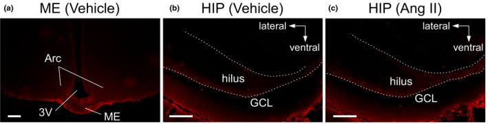
Methods: As a probe for accessibility, Evans blue dye (EB) that rapidly binds to albumin (Alb), which is impermeable to the BBB, was injected intravenously. To increase the vascular permeability of the BBB, a daily single administration of angiotensin II (Ang II) was applied intravenously for seven consecutive days.

Results: Fifteen minutes after the injection of EB, histological observation revealed that a number of neurons had entrapped and accumulated EB into their cell bodies in the hippocampal dentate gyrus in all rats. Of these, relatively large oval neurons (>15 µm) in the hilus and molecular layer showed parvalbumin immunopositivity, indicating they are GABAergic interneurons. The population of EB-accumulating neurons (approximately 10 µm) were localized in the inner margin of the granule cell layer, suggesting they were granule cells. However, the number of EB-positive neurons did not change in rats treated with Ang II compared with vehicle injection.

Conclusions: These findings suggest an intriguing possibility that blood-derived proteins such as hormones have access to hippocampal neurons constitutively in the absence of stimuli that increase the vascular permeability of the BBB in a physiologically normal state.

Keywords: Evans blue dye; albumin; blood-brain barrier; granule cell layer; hilus.


Conflict of interest statement

Dr. Mukuda, Dr. Koyama, Dr. Nakane, and Prof. Kaidoh declare no conflicts of interest.

Figures

Figure 1
Figure 1
Immunostaining of albumin (Alb) extravasated into the brain. Photomicrographs show the median eminence (ME) (a) and hippocampus (HIP) (b, c). (a, b) Brain tissue slices obtained from a rat given a single injection of vehicle (300 µl). (c) Hippocampal section from a rat injected with Ang II (10–5 M, 200 µl plus 100 µl of vehicle). White broken lines on the HIP indicate the hilar border of the granule cell layer (GCL) of the dentate gyrus. Scale bars, 200 µm. Arc, arcuate nucleus; 3V, third ventricle
Figure 2
Figure 2
Accumulation of Evans blue dye (EB) in cell bodies in the brain. Rats were given EB (200 µl, 20 mg/ml in vehicle) via an implanted atrial catheter 15 min before sacrifice. (a, b) Photomicrographs show the uptake of EB in cells of the Arc in a rat injected with vehicle. The enclosed area with a white line in (a) is magnified in (b). (c) Low magnification of the HIP obtained from a rat given Ang II. (d, e) High magnification of the hippocampal dentate gyrus from rats given vehicle (d) or Ang II (e). White broken lines on the HIP indicate the hilar border of the GCL of the dentate gyrus. Scale bars, 200 µm
Figure 3
Figure 3
A box‐and‐whisker diagram showing the number of EB‐incorporating cells in the rostral part of the hippocampal dentate gyrus (from the rostral tip to 1,200 µm caudal) in rats given vehicle or Ang II. Statistical comparison was performed using an unpaired t test
Figure 4
Figure 4
Accumulation of EB in hippocampal and hypothalamic cells at different time points after EB injection. Rats were given EB (200 µl, 20 mg/ml in vehicle) via an implanted atrial catheter 15 min (a) or 2 days (b) before sacrifice. Photomicrographs show the HIP (top), periventricular (PVN) (middle), and supraoptic (SON) (bottom) nuclei, in each left hemisphere. White broken lines in the HIP indicate the hilar border of the GCL. Scale bars, 100 µm. OC, optic chiasma
Figure 5
Figure 5
Neural staining in EB‐incorporating cells of the HIP and Arc. (a, b) EB‐incorporating cells were labeled with a green fluorescent Nissl stain in the HIP (a) and Arc (b). Top panels show cells incorporating EB (red). Middle panels show magnified areas enclosed with white squares in the top panels, which are merged with signals of the green fluorescent Nissl stain (green) and DAPI (blue). Bottom panels are the respective color images of EB, Nissl, and DAPI in the area enclosed with white squares in the middle panels. (c, d) EB‐incorporating cells were immunostained for glial fibrillary acidic protein (GFAP) in the HIP (c) and Arc (d). Top panels show GFAP‐immunoreactive astrocytes (green), together with the EB signal. Bottom panels are the respective color images of EB and GFAP in the areas enclosed with white squares in the top panels. White broken lines in the HIP indicate the hilar border of the GCL. Scale bars, 100 µm (top in a, b), 50 µm (middle in a, b and top in c, d), and 20 µm (bottom in a–d)
Figure 6
Figure 6
Immunohistochemistry for parvalbumin (PV) in the HIP. Top panels show EB‐incorporating neurons (red). Bottom panels indicate PV immunoreactivity (green) together with EB and DAPI (blue) signals in the areas enclosed with white squares in the top panels. White broken lines indicate the hilar border of the GCL. Arrows indicate neurons double‐positive to EB and PV. Arrowheads indicate neurons positive for EB but negative for PV. Scale bars, 100 µm (top), 20 µm (bottom). ML, molecular layer
Figure 7
Figure 7
Immunohistochemistry for neurogenic marker molecules in the HIP. (a, b) Photomicrographs showing signals of 5‐bromo‐2′‐deoxyuridine (BrdU, green) (a) and doublecortin (DCX, green) (b), together with EB. Rats were given BrdU (10 mg/kg bw in vehicle) via an implanted atrial catheter once a day for four consecutive days before sacrifice. White broken lines indicate the hilar border of the GCL. Bottom panels magnify the areas enclosed with white squares in the top panels. Cell nuclei were counterstained with DAPI (blue) in the study for DCX. Scale bars, 100 µm (top), 20 µm (bottom)

References

    1. Albanese, A. , & Bentivoglio, M. (1982). Retrograde fluorescent neuronal tracing combined with acetylcholinesterase histochemistry. Journal of Neuroscience Methods, 6, 121–127. 10.1016/0165-0270(82)90022-X - DOI - PubMed
    1. Alonso, A. , Reinz, E. , Fatar, M. , Hennerici, M. G. , & Meairs, S. (2011). Clearance of albumin following ultrasound‐induced blood‐brain barrier opening is mediated by glial but not neuronal cells. Brain Research, 1411, 9–16. 10.1016/j.brainres.2011.07.006 - DOI - PubMed
    1. Amaral, S. L. , Papanek, P. E. , & Greene, A. S. (2001). Angiotensin II and VEGF are involved in angiogenesis induced by short‐term exercise training. The American Journal of Physiology‐Heart and Circulatory Physiology, 281, H1163–H1169. 10.1152/ajpheart.2001.281.3.H1163 - DOI - PubMed
    1. Banks, W. A. , Tschöp, M. , Robinson, S. M. , & Heiman, M. L. (2002). Extent and direction of ghrelin transport across the blood‐brain barrier is determined by its unique primary structure. Journal of Pharmacology and Experimental Therapeutics, 302, 822–827. 10.1124/jpet.102.034827 - DOI - PubMed
    1. Biancardi, V. C. , Son, S. J. , Ahmadi, S. , Filosa, J. A. , & Stern, J. E. (2014). Circulating angiotensin II gains access to the hypothalamus and brain stem during hypertension via breakdown of the blood‐brain barrier. Hypertension, 63, 572–579. 10.1161/HYPERTENSIONAHA.113.01743 - DOI - PMC - PubMed

Publication types