. 2020 Jan 27;12(3):2777-2797.
doi: 10.18632/aging.102776. Epub 2020 Jan 27.

TP53/miR-34a-associated signaling targets SERPINE1 expression in human pancreatic cancer

Affiliations

TP53/miR-34a-associated signaling targets SERPINE1 expression in human pancreatic cancer

Shaw M Akula et al. Aging (Albany NY). .

Abstract

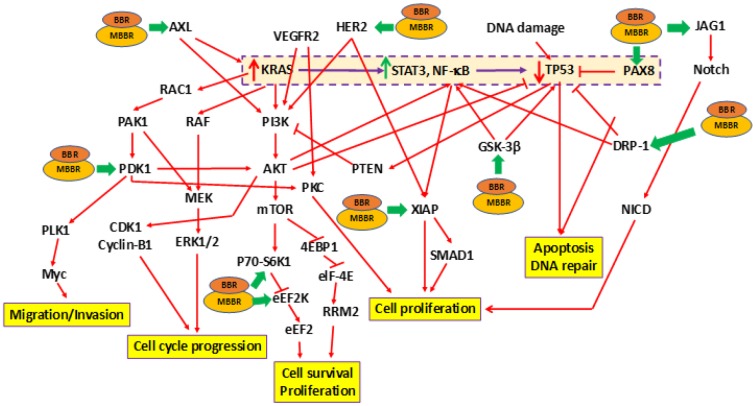
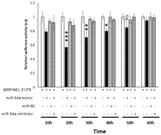
Pancreatic ductal adenocarcinoma (PDAC) is a disease of aging. The TP53 gene product regulates cell growth, aging, and cancer. To determine the important targets of TP53 in PDAC, we examined the expression of 440 proteins on a reverse phase protein array (RPPA) in PDAC-derived MIA-PaCa-2 cells which either had WT-TP53 or lacked WT-TP53. MIA-PaCa-2 cells have a TP53 mutation as well as mutant KRAS and represent a good in vitro model to study PDAC. RPPA analysis demonstrated expression of tumor promoting proteins in cells that lacked WT-TP53; and this feature could be reversed significantly when the cells were transfected with vector encoding WT-TP53 or treated with berberine or a modified berberine (BBR). Expression of miR-34a-associated signaling was elevated in cells expressing WT-TP53 compared to cells expressing mTP53. Results from in vivo studies using human PDAC specimens confirmed the in vitro results as the expression of miR-34a and associated signaling was significantly decreased in PDAC specimens compared to non-cancerous tissues. This study determined SERPINE1 as a miR-34a target with relevance to the biology of PDAC. Thus, we have identified a key target (SERPINE1) of the TP53/miR-34a axis that may serve as a potential biomarker for early detection of pancreatic cancer.

Keywords: Aging; PDAC; SERPINE1; TP53; cancer; miR-34a.


Conflict of interest statement

CONFLICTS OF INTEREST: The authors declare no conflicts of interest.

Figures

Figure 1
Figure 1
Changes in protein expression profile in MIA-PaCa-2 cells expressing pLXSN compared to WT-TP53. (A) Protein expression was assayed by RPPA. Proteins indicated in red and green denotes increased and decreased expression, respectively. Genes in red and green indicate tumor promoting and suppressor activities, respectively. (B) Schematic demonstrating cell signaling in MIA-PaCa-2+pLXSN cells promoting cell survival (in red) while significantly inhibiting apoptosis (in green).
Figure 2
Figure 2
Effects of treating MIA-PaCa-2 cells with BBR and NAX060 on cell division, proliferation, survival, migration, and apoptosis. A schematic depicting the effects of BBR and NAX060 on the N-RAS/TP53-associated signaling critical to PDAC development. The model is based on the fact that over-expression of mutated KRAS significantly enhances STAT3, NF-κB signaling which in turn lowers the TP53 expression (highlighted and boxed in dotted purple line). Green bold arrows denote inhibiting effects of BBR/MBBR on the signaling molecule.
Figure 3
Figure 3
miR-34a expression in MIA-PaCa-2+pLXSN cells. (A) qRT-PCR was conducted to determine the miR-34a expression in MIA-PaCa-2+WT-TP53 and MIA-PaCa-2+pLXSN cells. Briefly, approximately 500 ng of RNA was reverse transcribed in a 25 μl reaction volume using the All-in-one miRNA qRT-PCR detection kit (GeneCopoeia, Rockville, MD). The synthesized cDNAs were used in the PCR reaction. The expression levels of miR-34a were measured employing the SYBR green detection and specific forward primer for the mature miRNA sequence and the universal adaptor reverse primer (GeneCopoeia, USA). Two-tailed P value of 0.05 or less was considered statistically significant; ***p < 0.001. (B) The putative targets of miR-34a that were significantly altered in MIA-PaCa-2+pLXSN and MIA-PaCa-2+WT-TP53 cells when the cells were treated with BBR and MBBR. A select few of the miR-34a target proteins that were significantly altered by treatment of MIA-PaCa-2 cells with BBR and NAX060 are projected. The data represent average of three individual experiments. (C) qRT-PCR was conducted to determine the expression of miR-34a-target genes in MIA-PaCa2+pLXSN and MIA-PaCa-2+WT-TP53 cells. qRT-PCR was performed to monitor expression of the different miR-34a-putative target genes in untreated MIA-PaCa-2+pLXSN cells and MIA-PaCa-2 expressing WT-TP53 or those treated with BBR and MBBR, respectively, using specific primers and SYBR green detection as per standard protocols. Bars represent average ± s.d. of three individual experiments.
Figure 4
Figure 4
Expression profile of miR-34a in human PDAC samples. (A) miR34-a expression levels are lower in PDAC specimens compared to healthy pancreas controls. We compared the expression of miR-34a in 10 specimens in each group. Student t test was performed to compare groups. Two-tailed P value of 0.05 or less was considered statistically significant. ***p < 0.001. (B) qRT-PCR was conducted to determine the expression of miR-34a-target genes in human PDAC or healthy pancreas control specimens. Expression of miR-34a-target genes in human PDAC and healthy pancreas control specimens were detected by qRT-PCR using specific primers and SYBR green detection as per standard protocols. Bars represent average ± s.d. of three individual experiments. Two-tailed P value of 0.05 or less was considered statistically significant; ***p < 0.001.
Figure 5
Figure 5
miR-34a targets SERPINE1. (A, B) To determine the cytotoxic effect of miR-34a mimic and inhibitor, MIA-PaCa2+pLXSN cells were transfected with different concentrations of miR-34a mimic and inhibitor. At 24 h post transfection, MTT was added to each well and the absorption was measured. Percentage of cell death was monitored for miR-34a mimic (miR-mimic) (A) and miR-inhibitor (B) compared with 0.01% DMSO as control. (C, D) miR-34a mimic significantly decreased expression of SERPINE1 and PAI-1 in MIA-PaCa-2+pLXSN cells. MIA-PaCa-2+pLXSN cells were untransfected, mock transfected, or transfected with miR-34a mimic or miR-NC. At the end of 24h of incubation at 37°C, the cells were lysed, RNA extracted (panel C), cDNA synthesized, and SERPINE1 expression monitored by qRT-PCR. In another set of experiments, the cells were lysed were probed for PAI-1 expression by Western blotting (panel D). (E) The relative expression of SERPINE1 and miR-34a in MIA-PaCa-2 target cells was monitored by qRT-PCR. The expression was measured in terms of cycle threshold value (Ct) and normalized to expression of β-actin and snRNA RNU6B, respectively. The x-axis denotes the cell type and y-axis denotes fold change in expression of SERPINE1 and miR-34a. The R2 values for the miRNA expression are provided. (F) In another set of experiments, the above cells were lysed and probed for PAI-1 expression by Western blotting (panel F). Bars (AC, E) represent average ± s.d. of five individual experiments. Student t test was performed to compare groups. Two-tailed P value of 0.05 or less was considered statistically significant. **p,0.01; ***p < 0.001; NS-not significant.
Figure 6
Figure 6
miR-34a specifically binds and interact with SERPINE1. Luciferase activity in 293 cells transfected with Dual-luciferase vector encoding Gaussia Luciferase (GLuc) and secreted alkaline phosphatase (SEAP) with 3′UR of SERPINE1 placed downstream of Glu luciferase reporter (SERPINE1 3′UTR). 293 cells were either transfected with SERPINE1 3′UTR, co-transfected with SERPINE1 3′UTR and miR-34a mimic, co-transfected with SERPINE1 3′UTR and control mimic (miR-NC), or co-transfected with SERPINE1 3′UTR, miR-34a mimic and miR-34a inhibitor. GLuc activity was monitored at 10 h, 22 h, 30 h, 40h, 50 h, and 60 h post-transfection and was normalized to SEAP. Data is plotted as GLuc/SEAP ratio where the x-axis indicates the transfection and time points, and y-axis indicates the relative luciferase activity. Bars represent average ± s.d. of five individual experiments. Student t test was performed to compare groups. Two-tailed P value of 0.05 or less was considered statistically significant. *p < 0.05; **p,0.01; ***p < 0.001; NS-not significant.

References

    1. Higuera O, Ghanem I, Nasimi R, Prieto I, Koren L, Feliu J. Management of pancreatic cancer in the elderly. World J Gastroenterol. 2016; 22:764–75. 10.3748/wjg.v22.i2.764 - DOI - PMC - PubMed
    1. Idachaba S, Dada O, Abimbola O, Olayinka O, Uma A, Olunu E, Fakoya AO. A Review of Pancreatic Cancer: Epidemiology, Genetics, Screening, and Management. Open Access Maced J Med Sci. 2019; 7:663–71. 10.3889/oamjms.2019.104 - DOI - PMC - PubMed
    1. Patel N, Khorolsky C, Benipal B. Incidence of Pancreatic Adenocarcinoma in the United States from 2001 to 2015: A United States Cancer Statistics Analysis of 50 States. Cureus. 2018; 10:e3796. 10.7759/cureus.3796 - DOI - PMC - PubMed
    1. Jeune F, Coriat R, Prat F, Dousset B, Vaillant JC, Gaujoux S. Pancreatic cancer surgical management. Presse Med. 2019; 48:e147–58. 10.1016/j.lpm.2019.02.027 - DOI - PubMed
    1. Falzone L, Salomone S, Libra M. Evolution of Cancer Pharmacological Treatments at the Turn of the Third Millennium. Front Pharmacol. 2018; 9:1300. 10.3389/fphar.2018.01300 - DOI - PMC - PubMed

Publication types

MeSH terms