

Expression of degenerative markers in intervertebral discs of young and elderly asymptomatic individuals
- PMID: 31986181
- PMCID: PMC6984735
- DOI: 10.1371/journal.pone.0228155
Expression of degenerative markers in intervertebral discs of young and elderly asymptomatic individuals
Abstract
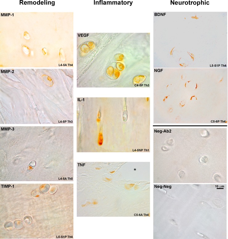
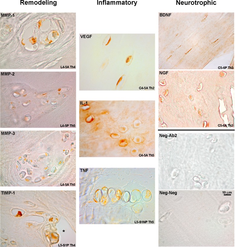
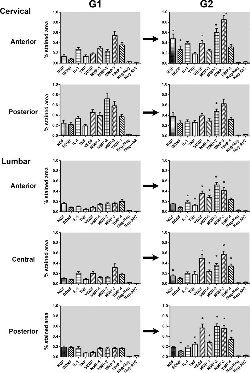
Intervertebral disc (IVD) degeneration is a remodeling process mediated by several growth factors and cytokines. This process has been extensively studied in vitro and with pathologic specimens obtained during surgery for scoliosis or back pain. However, the occurrence and temporal evolution of these molecules during normal aging, particularly in the cervical segment, is not known. Our objective was to study and compare the presence of putative mediators in the IVD of young (<35 years, G1) and elderly (>65 years, G2) presumably asymptomatic individuals. Thirty C4-5 and C5-6 discs and thirty L4-5 and L5-S1 discs per group were collected during the autopsy of individuals whose family members denied a history of neck or back pain. Discs were divided into anterior, central (lumbar only) and posterior sectors for analysis. Immunohistochemistry for TNF-α, IL-1β, VEGF, NGF-β, BDNF, TIMP-1, MMP-1, -2 and -3 was performed and reactivity compared between groups and sectors. All of these molecules were detected in every disc sector of both G1 and G2. Most statistical comparisons (25/45, 55.6%) revealed an increase in mediator expression in G2 in relation to G1. Regional differences in the expression of remodeling enzymes were rare; NGF-β and BDNF had slightly higher expression in the cervical segment of elderly individuals. A senescent profile with elevated VEGF, MMP-2 and MMP-3 was observed across most G2 disc regions and were generally elevated from G1. In conclusion, the mere presence of any of the studied molecules inside the IVD cannot be considered pathologic. Expression of remodeling enzymes and inflammatory mediators is relatively similar across different vertebral segments and disc regions leading to a common degenerated pattern, while neurotrophins have slightly higher expression in cervical discs. These findings support the concept that disc remodeling in different segments follows a similar pathway that can be potentially mediated to avoid structural failure.
Conflict of interest statement
The authors have declared that no competing interests exist.
Figures
Similar articles
-
Normal aging in human lumbar discs: An ultrastructural comparison.PLoS One. 2019 Jun 20;14(6):e0218121. doi: 10.1371/journal.pone.0218121. eCollection 2019. PLoS One. 2019. PMID: 31220091 Free PMC article.
-
Bone morphogenic protein-2 signaling in human disc degeneration and correlation to the Pfirrmann MRI grading system.Spine J. 2021 Jul;21(7):1205-1216. doi: 10.1016/j.spinee.2021.03.002. Epub 2021 Mar 5. Spine J. 2021. PMID: 33677096 Free PMC article.
-
Is coronal imbalance in degenerative lumbar scoliosis patients associated with the number of degenerated discs? A retrospective imaging cross-sectional study.BMC Musculoskelet Disord. 2023 May 25;24(1):414. doi: 10.1186/s12891-023-06558-9. BMC Musculoskelet Disord. 2023. PMID: 37231434 Free PMC article.
-
Structural and Ultrastructural Analysis of the Cervical Discs of Young and Elderly Humans.PLoS One. 2015 Oct 1;10(10):e0139283. doi: 10.1371/journal.pone.0139283. eCollection 2015. PLoS One. 2015. PMID: 26427056 Free PMC article.
-
The aging spine: the role of inflammatory mediators in intervertebral disc degeneration.Cell Mol Biol (Noisy-le-grand). 2007 May 30;53(5):4-18. Cell Mol Biol (Noisy-le-grand). 2007. PMID: 17543240 Review.
Cited by
-
Alterations of bovine nucleus pulposus cells with aging.Aging Cell. 2023 Aug;22(8):e13873. doi: 10.1111/acel.13873. Epub 2023 May 30. Aging Cell. 2023. PMID: 37254638 Free PMC article.
-
Preclinical ex-vivo Testing of Anti-inflammatory Drugs in a Bovine Intervertebral Degenerative Disc Model.Front Bioeng Biotechnol. 2020 Jun 10;8:583. doi: 10.3389/fbioe.2020.00583. eCollection 2020. Front Bioeng Biotechnol. 2020. PMID: 32587853 Free PMC article.
-
Low-dose celecoxib-loaded PCL fibers reverse intervertebral disc degeneration by up-regulating CHSY3 expression.J Nanobiotechnology. 2023 Mar 3;21(1):76. doi: 10.1186/s12951-023-01823-4. J Nanobiotechnology. 2023. PMID: 36864461 Free PMC article.
-
The interaction between CTGF and VEGF-A in the progression of intervertebral disc fibrosis.Afr Health Sci. 2024 Dec;24(4):276-285. doi: 10.4314/ahs.v24i4.36. Afr Health Sci. 2024. PMID: 40190527 Free PMC article.
-
Co-culture with Sirt1-overexpressed chondrocytes delays the nucleus pulposus cells degeneration.Cell Tissue Bank. 2022 Mar;23(1):57-66. doi: 10.1007/s10561-021-09912-0. Epub 2021 Mar 8. Cell Tissue Bank. 2022. PMID: 33683504
References
-
- Fardon DF, Milette PC. Nomenclature and classification of lumbar disc pathology. Recommendations of the Combined task Forces of the North American Spine Society, American Society of Spine Radiology, and American Society of Neuroradiology. Spine. 2001. March 1;26(5):E93–113. 10.1097/00007632-200103010-00006 - DOI - PubMed
-
- Koike Y, Uzuki M, Kokubun S, Sawai T. Angiogenesis and inflammatory cell infiltration in lumbar disc herniation. Spine. 2003. September 1;28(17):1928–33. 10.1097/01.BRS.0000083324.65405.AE - DOI - PubMed
MeSH terms
Substances
LinkOut - more resources
Full Text Sources
Research Materials
Miscellaneous
